5GVR | pdb_00005gvr

Crystal structure of the DDX41 DEAD domain in an apo closed form


Experimental Data Snapshot

  • Method: X-RAY DIFFRACTION
  • Resolution: 1.50 Å
  • R-Value Free: 
    0.208 (Depositor), 0.210 (DCC) 
  • R-Value Work: 
    0.181 (Depositor), 0.183 (DCC) 
  • R-Value Observed: 
    0.182 (Depositor) 

Starting Model: experimental
View more details

wwPDB Validation 3D Report Full Report

Validation slider image for 5GVR

This is version 1.1 of the entry. See complete history

Literature

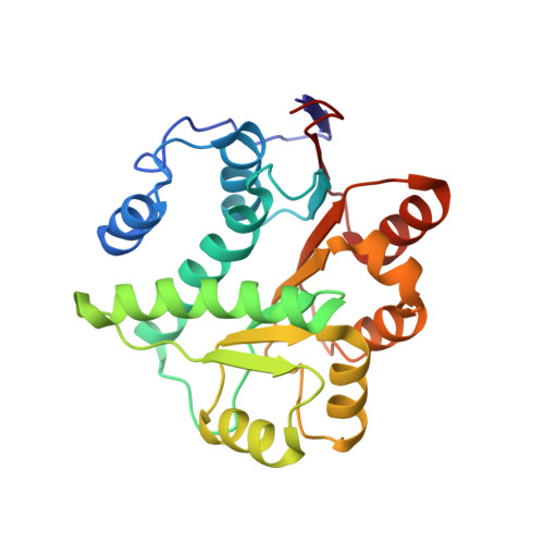
Structural and Functional Analysis of DDX41: a bispecific immune receptor for DNA and cyclic dinucleotide

Omura, H.Oikawa, D.Nakane, T.Kato, M.Ishii, R.Ishitani, R.Tokunaga, F.Nureki, O.

(2016) Sci Rep 6: 34756-34756

  • DOI: https://doi.org/10.1038/srep34756
  • Primary Citation Related Structures: 
    5GVR, 5GVS

  • PubMed Abstract: 

    In the innate immune system, pattern recognition receptors (PRRs) specifically recognize ligands derived from bacteria or viruses, to trigger the responsible downstream pathways. DEAD box protein 41 (DDX41) is an intracellular PRR that triggers the downstream pathway involving the adapter STING, the kinase TBK1, and the transcription factor IRF3, to activate the type I interferon response. DDX41 is unique in that it recognizes two different ligands; i.e., double-stranded DNA (dsDNA) and cyclic dinucleotides (CDN), via its DEAD domain. However, the structural basis for the ligand recognition by the DDX41 DEAD domain has remained elusive. Here, we report two crystal structures of the DDX41 DEAD domain in apo forms, at 1.5 and 2.2 Å resolutions. A comparison of the two crystal structures revealed the flexibility in the ATP binding site, suggesting its formation upon ATP binding. Structure-guided functional analyses in vitro and in vivo demonstrated the overlapped binding surface for dsDNA and CDN, which is distinct from the ATP-binding site. We propose that the structural rearrangement of the ATP binding site is crucial for the release of ADP, enabling the fast turnover of DDX41 for the dsDNA/CDN-induced STING activation pathway.


  • Organizational Affiliation
    • Department of Biophysics and Biochemistry, Graduate School of Science, The University of Tokyo, 2-11-16 Yayoi, Bunkyo-ku, Tokyo 113-0032, Japan.

Macromolecule Content 

  • Total Structure Weight: 26.31 kDa 
  • Atom Count: 1,949 
  • Modeled Residue Count: 234 
  • Deposited Residue Count: 234 
  • Unique protein chains: 1

Macromolecules

Find similar proteins by:|  3D Structure
Entity ID: 1
MoleculeChains  Sequence LengthOrganismDetailsImage
Probable ATP-dependent RNA helicase DDX41234Homo sapiensMutation(s): 0 
Gene Names: DDX41ABS
EC: 3.6.4.13
UniProt & NIH Common Fund Data Resources
Find proteins for Q9UJV9 (Homo sapiens)
Explore Q9UJV9 
Go to UniProtKB:  Q9UJV9
PHAROS:  Q9UJV9
GTEx:  ENSG00000183258 
Entity Groups
Sequence Clusters30% Identity50% Identity70% Identity90% Identity95% Identity100% Identity
UniProt GroupQ9UJV9
Sequence Annotations
Expand
Reference Sequence

Small Molecules

Ligands 1 Unique
IDChains Name / Formula / InChI Key2D Diagram3D Interactions
LMR

Query on LMR


Download:Ideal Coordinates CCD File
B [auth A](2S)-2-hydroxybutanedioic acid
C4 H6 O5
BJEPYKJPYRNKOW-REOHCLBHSA-N

Experimental Data & Validation

Experimental Data

  • Method: X-RAY DIFFRACTION
  • Resolution: 1.50 Å
  • R-Value Free:  0.208 (Depositor), 0.210 (DCC) 
  • R-Value Work:  0.181 (Depositor), 0.183 (DCC) 
  • R-Value Observed: 0.182 (Depositor) 
Space Group: P 32 2 1
Unit Cell:
Length ( Å )Angle ( ˚ )
a = 81.83α = 90
b = 81.83β = 90
c = 69.57γ = 120
Software Package:
Software NamePurpose
REFMACrefinement
PHASERphasing
REFMACmodel building
XDSdata scaling
XDSdata reduction
XDSdata processing
XDSdata extraction
XDSdata collection

Structure Validation

View Full Validation Report


Entry History 

Deposition Data

Revision History  (Full details and data files)

  • Version 1.0: 2016-10-19
    Type: Initial release
  • Version 1.1: 2023-11-08
    Changes: Data collection, Database references, Derived calculations, Refinement description