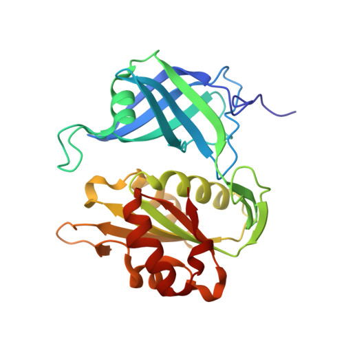
Distribution of valence electrons of the flavin cofactor in NADH-cytochrome b5 reductase.
Takaba, K., Takeda, K., Kosugi, M., Tamada, T., Miki, K.(2017) Sci Rep 7: 43162-43162
- PubMed: 28225078 Search on PubMedSearch on PubMed Central
- DOI: https://doi.org/10.1038/srep43162
- Primary Citation Related Structures:
5GV7, 5GV8 - PubMed Abstract:
Flavin compounds such as flavin adenine dinucleotide (FAD), flavin mononucleotide and riboflavin make up the active centers in flavoproteins that facilitate various oxidoreductive processes. The fine structural features of the hydrogens and valence electrons of the flavin molecules in the protein environment are critical to the functions of the flavoproteins. However, information on these features cannot be obtained from conventional protein X-ray analyses at ordinary resolution. Here we report the charge density analysis of a flavoenzyme, NADH-cytochrome b 5 reductase (b5R), at an ultra-high resolution of 0.78 Å. Valence electrons on the FAD cofactor as well as the peptide portion, which are clearly visualized even after the conventional refinement, are analyzed by the multipolar atomic model refinement. The topological analysis for the determined electron density reveals the valence electronic structure of the isoalloxazine ring of FAD and hydrogen-bonding interactions with the protein environment. The tetrahedral electronic distribution around the N5 atom of FAD in b5R is stabilized by hydrogen bonding with C α H of Tyr65 and amide-H of Thr66. The hydrogen bonding network leads to His49 composing the cytochrome b 5 -binding site via non-classical hydrogen bonds between N5 of FAD and C α H of Tyr65 and O of Tyr65 and C β H of His49.
- Department of Chemistry, Graduate School of Science, Kyoto University, Sakyo-ku, Kyoto 606-8502, Japan.
Organizational Affiliation: