5G5G

Escherichia coli Periplasmic Aldehyde Oxidase


Experimental Data Snapshot

  • Method: X-RAY DIFFRACTION
  • Resolution: 1.70 Å
  • R-Value Free: 0.167 
  • R-Value Work: 0.138 
  • R-Value Observed: 0.140 

wwPDB Validation   3D Report Full Report


Ligand Structure Quality Assessment 


This is version 1.2 of the entry. See complete history


Literature

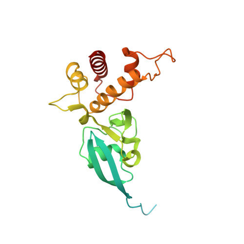
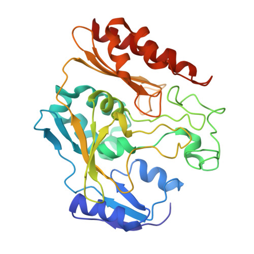
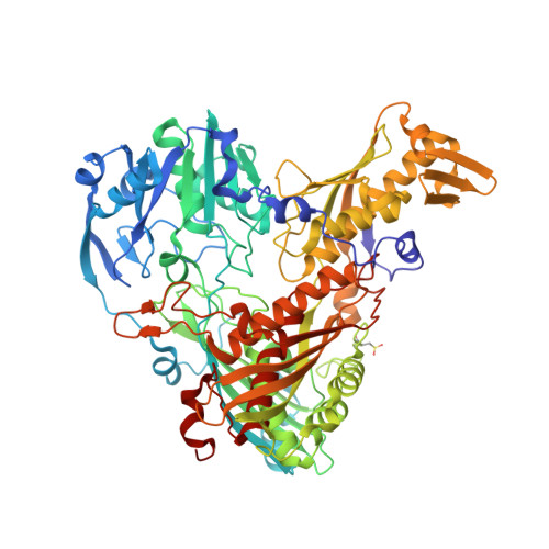
The Escherichia Coli Periplasmic Aldehyde Oxidoreductase is an Exceptional Member of the Xanthine Oxidase Family of Molybdoenzymes.

Correia, M.A.Otrelo-Cardoso, A.R.Schwuchow, V.Sigfridsson Clauss, K.G.Haumann, M.Romao, M.J.Leimkuhler, S.Santos-Silva, T.

(2016) ACS Chem Biol 11: 2923

  • DOI: https://doi.org/10.1021/acschembio.6b00572
  • Primary Citation of Related Structures:  
    5G5G, 5G5H

  • PubMed Abstract: 
  • The xanthine oxidase (XO) family comprises molybdenum-dependent enzymes that usually form homodimers (or dimers of heterodimers/trimers) organized in three domains that harbor two [2Fe-2S] clusters, one FAD, and a Mo cofactor. In this work, we crystallized an unusual member of the family, the periplasmic aldehyde oxidoreductase PaoABC from Escherichia coli ...

    The xanthine oxidase (XO) family comprises molybdenum-dependent enzymes that usually form homodimers (or dimers of heterodimers/trimers) organized in three domains that harbor two [2Fe-2S] clusters, one FAD, and a Mo cofactor. In this work, we crystallized an unusual member of the family, the periplasmic aldehyde oxidoreductase PaoABC from Escherichia coli. This is the first example of an E. coli protein containing a molybdopterin-cytosine-dinucleotide cofactor and is the only heterotrimer of the XO family so far structurally characterized. The crystal structure revealed the presence of an unexpected [4Fe-4S] cluster, anchored to an additional 40 residues subdomain. According to phylogenetic analysis, proteins containing this cluster are widely spread in many bacteria phyla, putatively through repeated gene transfer events. The active site of PaoABC is highly exposed to the surface with no aromatic residues and an arginine (PaoC-R440) making a direct interaction with PaoC-E692, which acts as a base catalyst. In order to understand the importance of R440, kinetic assays were carried out, and the crystal structure of the PaoC-R440H variant was also determined.


    Organizational Affiliation

    UCIBIO/Requimte, Departamento de Química, Faculdade de Ciências e Tecnologia, Universidade Nova de Lisboa , 2829-516 Caparica, Portugal.


Macromolecules
Find similar proteins by:  (by identity cutoff)  |  3D Structure
Entity ID: 1
MoleculeChainsSequence LengthOrganismDetailsImage
PUTATIVE XANTHINE DEHYDROGENASE YAGT IRON-SULFUR-BINDING SUBUNIT229Escherichia coli K-12Mutation(s): 0 
Gene Names: paoAyagTb0286JW0280
EC: 1.2.99.6
UniProt
Find proteins for P77165 (Escherichia coli (strain K12))
Explore P77165 
Go to UniProtKB:  P77165
Entity Groups  
Sequence Clusters30% Identity50% Identity70% Identity90% Identity95% Identity100% Identity
UniProt GroupP77165
Protein Feature View
Expand
  • Reference Sequence
Find similar proteins by:  (by identity cutoff)  |  3D Structure
Entity ID: 2
MoleculeChainsSequence LengthOrganismDetailsImage
PUTATIVE XANTHINE DEHYDROGENASE YAGR MOLYBDENUM-BINDING SU SUBUNIT318Escherichia coli K-12Mutation(s): 0 
Gene Names: paoByagSb0285JW0279
EC: 1.2.99.6
UniProt
Find proteins for P77324 (Escherichia coli (strain K12))
Explore P77324 
Go to UniProtKB:  P77324
Entity Groups  
Sequence Clusters30% Identity50% Identity70% Identity90% Identity95% Identity100% Identity
UniProt GroupP77324
Protein Feature View
Expand
  • Reference Sequence
Find similar proteins by:  (by identity cutoff)  |  3D Structure
Entity ID: 3
MoleculeChainsSequence LengthOrganismDetailsImage
PUTATIVE XANTHINE DEHYDROGENASE YAGS FAD-BINDING SUBUNIT732Escherichia coli K-12Mutation(s): 0 
Gene Names: paoCyagRb0284JW0278
EC: 1.2.99.6
UniProt
Find proteins for P77489 (Escherichia coli (strain K12))
Explore P77489 
Go to UniProtKB:  P77489
Entity Groups  
Sequence Clusters30% Identity50% Identity70% Identity90% Identity95% Identity100% Identity
UniProt GroupP77489
Protein Feature View
Expand
  • Reference Sequence
Small Molecules
Ligands 9 Unique
IDChainsName / Formula / InChI Key2D Diagram3D Interactions
FAD
Query on FAD

Download Ideal Coordinates CCD File 
O [auth B]FLAVIN-ADENINE DINUCLEOTIDE
C27 H33 N9 O15 P2
VWWQXMAJTJZDQX-UYBVJOGSSA-N
 Ligand Interaction
MCN
Query on MCN

Download Ideal Coordinates CCD File 
Y [auth C]PTERIN CYTOSINE DINUCLEOTIDE
C19 H22 N8 O13 P2 S2
RBWYFPNWTRZKKZ-LOIMWUFNSA-N
 Ligand Interaction
SF4
Query on SF4

Download Ideal Coordinates CCD File 
N [auth B]IRON/SULFUR CLUSTER
Fe4 S4
LJBDFODJNLIPKO-VKOJMFJBAC
 Ligand Interaction
FES
Query on FES

Download Ideal Coordinates CCD File 
D [auth A],
E [auth A]
FE2/S2 (INORGANIC) CLUSTER
Fe2 S2
NIXDOXVAJZFRNF-UHFFFAOYSA-N
 Ligand Interaction
MOS
Query on MOS

Download Ideal Coordinates CCD File 
Z [auth C]DIOXOTHIOMOLYBDENUM(VI) ION
H Mo O2 S
BDSRWPHSAKXXRG-UHFFFAOYSA-M
 Ligand Interaction
IOD
Query on IOD

Download Ideal Coordinates CCD File 
AA [auth C]
BA [auth C]
CA [auth C]
F [auth A]
M [auth A]
AA [auth C],
BA [auth C],
CA [auth C],
F [auth A],
M [auth A],
P [auth B],
W [auth B]
IODIDE ION
I
XMBWDFGMSWQBCA-UHFFFAOYSA-M
 Ligand Interaction
GOL
Query on GOL

Download Ideal Coordinates CCD File 
JA [auth C]
K [auth A]
KA [auth C]
L [auth A]
U [auth B]
JA [auth C],
K [auth A],
KA [auth C],
L [auth A],
U [auth B],
V [auth B]
GLYCEROL
C3 H8 O3
PEDCQBHIVMGVHV-UHFFFAOYSA-N
 Ligand Interaction
ACT
Query on ACT

Download Ideal Coordinates CCD File 
HA [auth C],
IA [auth C],
J [auth A],
T [auth B]
ACETATE ION
C2 H3 O2
QTBSBXVTEAMEQO-UHFFFAOYSA-M
 Ligand Interaction
CL
Query on CL

Download Ideal Coordinates CCD File 
DA [auth C]
EA [auth C]
FA [auth C]
G [auth A]
GA [auth C]
DA [auth C],
EA [auth C],
FA [auth C],
G [auth A],
GA [auth C],
H [auth A],
I [auth A],
LA [auth C],
Q [auth B],
R [auth B],
S [auth B],
X [auth B]
CHLORIDE ION
Cl
VEXZGXHMUGYJMC-UHFFFAOYSA-M
 Ligand Interaction
Modified Residues  1 Unique
IDChainsTypeFormula2D DiagramParent
CSD
Query on CSD
C
L-PEPTIDE LINKINGC3 H7 N O4 SCYS
Experimental Data & Validation

Experimental Data

  • Method: X-RAY DIFFRACTION
  • Resolution: 1.70 Å
  • R-Value Free: 0.167 
  • R-Value Work: 0.138 
  • R-Value Observed: 0.140 
  • Space Group: C 1 2 1
Unit Cell:
Length ( Å )Angle ( ˚ )
a = 109.681α = 90
b = 78.342β = 99.69
c = 151.909γ = 90
Software Package:
Software NamePurpose
REFMACrefinement
XDSdata reduction
Aimlessdata scaling
PHASERphasing

Structure Validation

View Full Validation Report


Ligand Structure Quality Assessment 


Entry History 

Deposition Data

Revision History  (Full details and data files)

  • Version 1.0: 2016-09-28
    Type: Initial release
  • Version 1.1: 2016-11-09
    Changes: Database references
  • Version 1.2: 2017-09-13
    Changes: Structure summary