5G5D

Crystal Structure of the CohScaC2-XDocCipA type II complex from Clostridium thermocellum


Experimental Data Snapshot

  • Method: X-RAY DIFFRACTION
  • Resolution: 3.00 Å
  • R-Value Free: 0.303 
  • R-Value Work: 0.256 
  • R-Value Observed: 0.261 

wwPDB Validation   3D Report Full Report


This is version 1.1 of the entry. See complete history


Literature

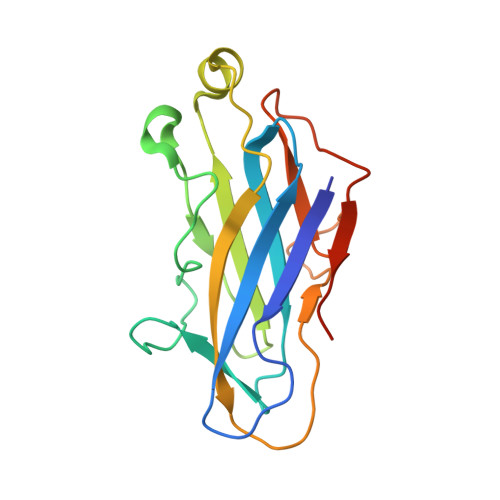
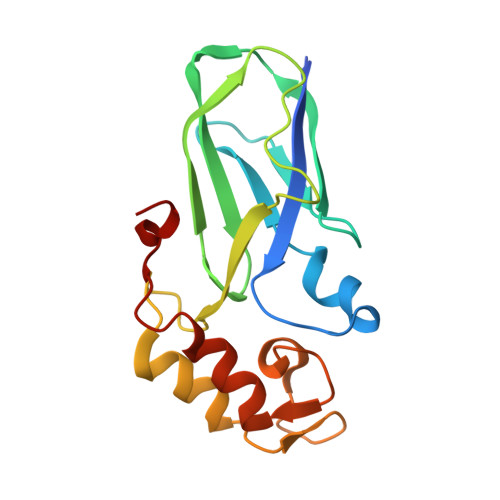
Diverse specificity of cellulosome attachment to the bacterial cell surface.

Bras, J.L.Pinheiro, B.A.Cameron, K.Cuskin, F.Viegas, A.Najmudin, S.Bule, P.Pires, V.M.Romao, M.J.Bayer, E.A.Spencer, H.L.Smith, S.Gilbert, H.J.Alves, V.D.Carvalho, A.L.Fontes, C.M.

(2016) Sci Rep 6: 38292-38292

  • DOI: https://doi.org/10.1038/srep38292
  • Primary Citation of Related Structures:  
    5G5D, 5K39, 5M0Y

  • PubMed Abstract: 

    During the course of evolution, the cellulosome, one of Nature's most intricate multi-enzyme complexes, has been continuously fine-tuned to efficiently deconstruct recalcitrant carbohydrates. To facilitate the uptake of released sugars, anaerobic bacteria use highly ordered protein-protein interactions to recruit these nanomachines to the cell surface. Dockerin modules located within a non-catalytic macromolecular scaffold, whose primary role is to assemble cellulosomal enzymatic subunits, bind cohesin modules of cell envelope proteins, thereby anchoring the cellulosome onto the bacterial cell. Here we have elucidated the unique molecular mechanisms used by anaerobic bacteria for cellulosome cellular attachment. The structure and biochemical analysis of five cohesin-dockerin complexes revealed that cell surface dockerins contain two cohesin-binding interfaces, which can present different or identical specificities. In contrast to the current static model, we propose that dockerins utilize multivalent modes of cohesin recognition to recruit cellulosomes to the cell surface, a mechanism that maximises substrate access while facilitating complex assembly.


  • Organizational Affiliation

    Centro Interdisciplinar de Investigação em Sanidade Animal, Faculdade de Medicina Veterinária, Universidade de Lisboa, 1300-477 Lisboa, Portugal.


Macromolecules
Find similar proteins by:  (by identity cutoff)  |  3D Structure
Entity ID: 1
MoleculeChains Sequence LengthOrganismDetailsImage
CELLULOSOME ANCHORING PROTEIN COHESIN REGION171Acetivibrio thermocellus AD2Mutation(s): 0 
UniProt
Find proteins for Q06853 (Acetivibrio thermocellus (strain ATCC 27405 / DSM 1237 / JCM 9322 / NBRC 103400 / NCIMB 10682 / NRRL B-4536 / VPI 7372))
Explore Q06853 
Go to UniProtKB:  Q06853
Entity Groups  
Sequence Clusters30% Identity50% Identity70% Identity90% Identity95% Identity100% Identity
UniProt GroupQ06853
Sequence Annotations
Expand
  • Reference Sequence
Find similar proteins by:  (by identity cutoff)  |  3D Structure
Entity ID: 2
MoleculeChains Sequence LengthOrganismDetailsImage
CELLULOSOMAL-SCAFFOLDING PROTEIN A164Acetivibrio thermocellus ATCC 27405Mutation(s): 0 
UniProt
Find proteins for Q06851 (Acetivibrio thermocellus (strain ATCC 27405 / DSM 1237 / JCM 9322 / NBRC 103400 / NCIMB 10682 / NRRL B-4536 / VPI 7372))
Explore Q06851 
Go to UniProtKB:  Q06851
Entity Groups  
Sequence Clusters30% Identity50% Identity70% Identity90% Identity95% Identity100% Identity
UniProt GroupQ06851
Sequence Annotations
Expand
  • Reference Sequence
Experimental Data & Validation

Experimental Data

  • Method: X-RAY DIFFRACTION
  • Resolution: 3.00 Å
  • R-Value Free: 0.303 
  • R-Value Work: 0.256 
  • R-Value Observed: 0.261 
  • Space Group: I 2 2 2
Unit Cell:
Length ( Å )Angle ( ˚ )
a = 52.36α = 90
b = 125.805β = 90
c = 130.467γ = 90
Software Package:
Software NamePurpose
REFMACrefinement
XDSdata reduction
SCALAdata scaling
PHASERphasing

Structure Validation

View Full Validation Report


Entry History 

Deposition Data

Revision History  (Full details and data files)

  • Version 1.0: 2017-04-05
    Type: Initial release
  • Version 1.1: 2024-01-10
    Changes: Data collection, Database references, Derived calculations, Other, Refinement description