5G1S | pdb_00005g1s

Open conformation of Francisella tularensis ClpP at 1.7 A


Experimental Data Snapshot

  • Method: X-RAY DIFFRACTION
  • Resolution: 1.70 Å
  • R-Value Free: 
    0.242 (Depositor), 0.246 (DCC) 
  • R-Value Work: 
    0.204 (Depositor), 0.212 (DCC) 
  • R-Value Observed: 
    0.206 (Depositor) 

Starting Model: other
View more details

wwPDB Validation 3D Report Full Report

Validation slider image for 5G1S

This is version 1.4 of the entry. See complete history

Literature

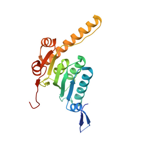
Open and compressed conformations of Francisella tularensis ClpP.

Diaz-Saez, L.Pankov, G.Hunter, W.N.

(2017) Proteins 85: 188-194

  • DOI: https://doi.org/10.1002/prot.25197
  • Primary Citation Related Structures: 
    5G1Q, 5G1R, 5G1S

  • PubMed Abstract: 

    Caseinolytic proteases are large oligomeric assemblies responsible for maintaining protein homeostasis in bacteria and in so doing influence a wide range of biological processes. The functional assembly involves three chaperones together with the oligomeric caseinolytic protease catalytic subunit P (ClpP). This protease represents a potential target for therapeutic intervention in pathogenic bacteria. Here, we detail an efficient protocol for production of recombinant ClpP from Francisella tularensis, and the structural characterization of three crystal forms which grow under similar conditions. One crystal form reveals a compressed state of the ClpP tetradecamer and two forms an open state. A comparison of the two types of structure infers that differences at the enzyme active site result from a conformational change involving a highly localized disorder-order transition of a β-strand α-helix combination. This transition occurs at a subunit-subunit interface. Our study may now underpin future efforts in a structure-based approach to target ClpP for inhibitor or activator development. Proteins 2016; 85:188-194. © 2016 Wiley Periodicals, Inc.


  • Organizational Affiliation
    • Division of Biological Chemistry and Drug Discovery, School of Life Sciences, University of Dundee, Dundee, United Kingdom.

Macromolecule Content 

  • Total Structure Weight: 470.75 kDa 
  • Atom Count: 37,499 
  • Modeled Residue Count: 3,966 
  • Deposited Residue Count: 4,221 
  • Unique protein chains: 1

Macromolecules

Find similar proteins by:|  3D Structure
Entity ID: 1
MoleculeChains  Sequence LengthOrganismDetailsImage
ATP-dependent Clp protease proteolytic subunit
A, B, C, D, E
A, B, C, D, E, F, G, H, I, J, K, L, M, N, O, P, Q, R, S, T, U
201Francisella tularensisMutation(s): 0 
Gene Names: clpPDR86_1343
EC: 3.4.21.92
UniProt
Find proteins for Q5NH47 (Francisella tularensis subsp. tularensis (strain SCHU S4 / Schu 4))
Explore Q5NH47 
Go to UniProtKB:  Q5NH47
Entity Groups
Sequence Clusters30% Identity50% Identity70% Identity90% Identity95% Identity100% Identity
UniProt GroupQ5NH47
Sequence Annotations
Expand
Reference Sequence

Small Molecules

Ligands 3 Unique
IDChains Name / Formula / InChI Key2D Diagram3D Interactions
MRD

Query on MRD


Download:Ideal Coordinates CCD File
AA [auth C]
BA [auth C]
DB [auth N]
EA [auth D]
FB [auth O]
AA [auth C],
BA [auth C],
DB [auth N],
EA [auth D],
FB [auth O],
HB [auth P],
JB [auth Q],
KA [auth G],
NB [auth S],
PA [auth I],
QB [auth T],
RB [auth T],
TA [auth K],
TB [auth U],
UA [auth K],
V [auth A],
VA [auth K],
XA [auth L]
(4R)-2-METHYLPENTANE-2,4-DIOL
C6 H14 O2
SVTBMSDMJJWYQN-RXMQYKEDSA-N
MPD

Query on MPD


Download:Ideal Coordinates CCD File
AB [auth M]
CB [auth N]
FA [auth E]
HA [auth F]
IA [auth F]
AB [auth M],
CB [auth N],
FA [auth E],
HA [auth F],
IA [auth F],
LA [auth G],
MB [auth S],
OA [auth I],
PB [auth T],
RA [auth J],
UB [auth U],
X [auth B],
YA [auth L],
Z [auth C]
(4S)-2-METHYL-2,4-PENTANEDIOL
C6 H14 O2
SVTBMSDMJJWYQN-YFKPBYRVSA-N
ACT

Query on ACT


Download:Ideal Coordinates CCD File
BB [auth M]
CA [auth C]
DA [auth D]
EB [auth N]
GA [auth E]
BB [auth M],
CA [auth C],
DA [auth D],
EB [auth N],
GA [auth E],
GB [auth O],
IB [auth P],
JA [auth F],
KB [auth Q],
LB [auth R],
MA [auth G],
NA [auth H],
OB [auth S],
QA [auth I],
SA [auth J],
SB [auth T],
VB [auth U],
W [auth A],
WA [auth K],
Y [auth B],
ZA [auth L]
ACETATE ION
C2 H3 O2
QTBSBXVTEAMEQO-UHFFFAOYSA-M

Experimental Data & Validation

Experimental Data

  • Method: X-RAY DIFFRACTION
  • Resolution: 1.70 Å
  • R-Value Free:  0.242 (Depositor), 0.246 (DCC) 
  • R-Value Work:  0.204 (Depositor), 0.212 (DCC) 
  • R-Value Observed: 0.206 (Depositor) 
Space Group: P 2 21 21
Unit Cell:
Length ( Å )Angle ( ˚ )
a = 98.226α = 90
b = 127.924β = 90
c = 353.843γ = 90
Software Package:
Software NamePurpose
REFMACrefinement
XDSdata reduction
Aimlessdata scaling
PHASERphasing

Structure Validation

View Full Validation Report


Entry History 

Deposition Data

Revision History  (Full details and data files)

  • Version 1.0: 2016-10-19
    Type: Initial release
  • Version 1.1: 2017-03-15
    Changes: Database references
  • Version 1.2: 2019-03-06
    Changes: Advisory, Data collection, Experimental preparation
  • Version 1.3: 2019-05-08
    Changes: Data collection, Experimental preparation
  • Version 1.4: 2024-05-01
    Changes: Advisory, Data collection, Database references, Derived calculations, Other, Refinement description