


The Structure of the 6-Carboxyhexanoate-Coa Ligase from Bacillus Subtilis
Wang, M., Moynie, L., Campopiano, D.J., Naismith, J.H.To be published.
Experimental Data Snapshot
Starting Model: experimental
View more details
Entity ID: 1 | |||||
|---|---|---|---|---|---|
| Molecule | Chains | Sequence Length | Organism | Details | Image |
| 6-CARBOXYHEXANOATE-COA LIGASE | 260 | Bacillus subtilis | Mutation(s): 0 EC: 6.2.1.14 | 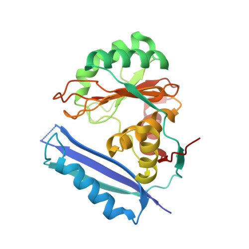 | |
UniProt | |||||
Entity Groups | |||||
| Sequence Clusters | 30% Identity50% Identity70% Identity90% Identity95% Identity100% Identity | ||||
| UniProt Group | P53559 | ||||
Sequence AnnotationsExpand | |||||
Reference Sequence | |||||
| Ligands 3 Unique | |||||
|---|---|---|---|---|---|
| ID | Chains | Name / Formula / InChI Key | 2D Diagram | 3D Interactions | |
| COA Download:Ideal Coordinates CCD File | E [auth B] | COENZYME A C21 H36 N7 O16 P3 S RGJOEKWQDUBAIZ-IBOSZNHHSA-N |  | ||
| 2PE Download:Ideal Coordinates CCD File | D [auth A] | NONAETHYLENE GLYCOL C18 H38 O10 YZUUTMGDONTGTN-UHFFFAOYSA-N |  | ||
| PML Download:Ideal Coordinates CCD File | C [auth A], F [auth B] | PIMELIC ACID C7 H12 O4 WLJVNTCWHIRURA-UHFFFAOYSA-N |  | ||
| Length ( Å ) | Angle ( ˚ ) |
|---|---|
| a = 50.59 | α = 90 |
| b = 78.36 | β = 90 |
| c = 166.43 | γ = 90 |
| Software Name | Purpose |
|---|---|
| REFMAC | refinement |
| xia2 | data reduction |
| XDS | data reduction |
| xia2 | data scaling |
| XSCALE | data scaling |
| PHASER | phasing |


