5FIA | pdb_00005fia

Structure of the effector protein LpiR1 (Lpg0634) from Legionella pneumophila


Experimental Data Snapshot

  • Method: X-RAY DIFFRACTION
  • Resolution: 1.75 Å
  • R-Value Free: 
    0.190 (Depositor), 0.199 (DCC) 
  • R-Value Work: 
    0.162 (Depositor) 
  • R-Value Observed: 
    0.163 (Depositor) 

wwPDB Validation 3D Report Full Report

Validation slider image for 5FIA

This is version 1.3 of the entry. See complete history

Literature

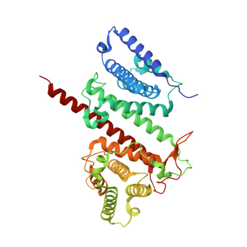
Structural and Functional Investigations of the Effector Protein LpiR1 from Legionella pneumophila.

Beyrakhova, K.A.van Straaten, K.Li, L.Boniecki, M.T.Anderson, D.H.Cygler, M.

(2016) J Biological Chem 291: 15767-15777

  • DOI: https://doi.org/10.1074/jbc.M115.708701
  • Primary Citation Related Structures: 
    5FIA, 5JG4

  • PubMed Abstract: 

    Legionella pneumophila is a causative agent of a severe pneumonia, known as Legionnaires' disease. Legionella pathogenicity is mediated by specific virulence factors, called bacterial effectors, which are injected into the invaded host cell by the bacterial type IV secretion system. Bacterial effectors are involved in complex interactions with the components of the host cell immune and signaling pathways, which eventually lead to bacterial survival and replication inside the mammalian cell. Structural and functional studies of bacterial effectors are, therefore, crucial for elucidating the mechanisms of Legionella virulence. Here we describe the crystal structure of the LpiR1 (Lpg0634) effector protein and investigate the effects of its overexpression in mammalian cells. LpiR1 is an α-helical protein that consists of two similar domains aligned in an antiparallel fashion. The hydrophilic cleft between the domains might serve as a binding site for a potential host cell interaction partner. LpiR1 binds the phosphate group at a conserved site and is stabilized by Mn(2+), Ca(2+), or Mg(2+) ions. When overexpressed in mammalian cells, a GFP-LpiR1 fusion protein is localized in the cytoplasm. Intracellular signaling antibody array analysis revealed small changes in the phosphorylation state of several components of the Akt signaling pathway in HEK293T cells overexpressing LpiR1.


  • Organizational Affiliation
    • From the Department of Biochemistry and.

Macromolecule Content 

  • Total Structure Weight: 91.95 kDa 
  • Atom Count: 6,939 
  • Modeled Residue Count: 787 
  • Deposited Residue Count: 804 
  • Unique protein chains: 1

Macromolecules

Find similar proteins by:|  3D Structure
Entity ID: 1
MoleculeChains  Sequence LengthOrganismDetailsImage
LpiR1
A, B
402Legionella pneumophila subsp. pneumophila str. Philadelphia 1Mutation(s): 0 
Gene Names: lpymg_00644
UniProt
Find proteins for Q5ZXU7 (Legionella pneumophila subsp. pneumophila (strain Philadelphia 1 / ATCC 33152 / DSM 7513))
Explore Q5ZXU7 
Go to UniProtKB:  Q5ZXU7
Entity Groups
Sequence Clusters30% Identity50% Identity70% Identity90% Identity95% Identity100% Identity
UniProt GroupQ5ZXU7
Sequence Annotations
Expand
Reference Sequence

Experimental Data & Validation

Experimental Data

  • Method: X-RAY DIFFRACTION
  • Resolution: 1.75 Å
  • R-Value Free:  0.190 (Depositor), 0.199 (DCC) 
  • R-Value Work:  0.162 (Depositor) 
  • R-Value Observed: 0.163 (Depositor) 
Space Group: P 1 21 1
Unit Cell:
Length ( Å )Angle ( ˚ )
a = 81.42α = 90
b = 71.82β = 111.96
c = 94.18γ = 90
Software Package:
Software NamePurpose
PHENIXrefinement
XDSdata reduction
XDSdata scaling
HKL-3000phasing

Structure Validation

View Full Validation Report


Entry History 

& Funding Information

Deposition Data


Funding OrganizationLocationGrant Number
Canadian Institutes of Health ReasearchCanadaMOP-48370

Revision History  (Full details and data files)

  • Version 1.0: 2016-01-27
    Type: Initial release
  • Version 1.1: 2016-06-08
    Changes: Database references
  • Version 1.2: 2016-08-03
    Changes: Database references
  • Version 1.3: 2024-03-06
    Changes: Data collection, Database references, Derived calculations