5FCW

HDAC8 Complexed with a Hydroxamic Acid


Experimental Data Snapshot

  • Method: X-RAY DIFFRACTION
  • Resolution: 1.98 Å
  • R-Value Free: 0.216 
  • R-Value Work: 0.172 
  • R-Value Observed: 0.174 

wwPDB Validation   3D Report Full Report


Ligand Structure Quality Assessment 


This is version 1.1 of the entry. See complete history


Literature

Structure of 'linkerless' hydroxamic acid inhibitor-HDAC8 complex confirms the formation of an isoform-specific subpocket.

Tabackman, A.A.Frankson, R.Marsan, E.S.Perry, K.Cole, K.E.

(2016) J Struct Biol 195: 373-378

  • DOI: https://doi.org/10.1016/j.jsb.2016.06.023
  • Primary Citation of Related Structures:  
    5FCW

  • PubMed Abstract: 

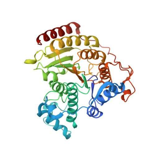
    Histone deacetylases (HDACs) catalyze the hydrolysis of acetylated lysine side chains in histone and non-histone proteins, and play a critical role in the regulation of many biological processes, including cell differentiation, proliferation, senescence, and apoptosis. Aberrant HDAC activity is associated with cancer, making these enzymes important targets for drug design. In general, HDAC inhibitors (HDACi) block the proliferation of tumor cells by inducing cell differentiation, cell cycle arrest, and/or apoptosis, and comprise some of the leading therapies in cancer treatments. To date, four HDACi have been FDA approved for the treatment of cancers: suberoylanilide hydroxamic acid (SAHA, Vorinostat, Zolinza®), romidepsin (FK228, Istodax®), belinostat (Beleodaq®), and panobinostat (Farydak®). Most current inhibitors are pan-HDACi, and non-selectively target a number of HDAC isoforms. Six previously reported HDACi were rationally designed, however, to target a unique sub-pocket found only in HDAC8. While these inhibitors were indeed potent against HDAC8, and even demonstrated specificity for HDAC8 over HDACs 1 and 6, there were no structural data to confirm the mode of binding. Here we report the X-ray crystal structure of Compound 6 complexed with HDAC8 to 1.98Å resolution. We also describe the use of molecular docking studies to explore the binding interactions of the other 5 related HDACi. Our studies confirm that the HDACi induce the formation of and bind in the HDAC8-specific subpocket, offering insights into isoform-specific inhibition.


  • Organizational Affiliation

    Department of Molecular Biology and Chemistry, Christopher Newport University, 1 Avenue of the Arts, Newport News, VA 23606, USA.


Macromolecules
Find similar proteins by:  (by identity cutoff)  |  3D Structure
Entity ID: 1
MoleculeChains Sequence LengthOrganismDetailsImage
Histone deacetylase 8
A, B
383Homo sapiensMutation(s): 0 
Gene Names: HDAC8HDACL1CDA07
EC: 3.5.1.98
UniProt & NIH Common Fund Data Resources
Find proteins for Q9BY41 (Homo sapiens)
Explore Q9BY41 
Go to UniProtKB:  Q9BY41
PHAROS:  Q9BY41
GTEx:  ENSG00000147099 
Entity Groups  
Sequence Clusters30% Identity50% Identity70% Identity90% Identity95% Identity100% Identity
UniProt GroupQ9BY41
Sequence Annotations
Expand
  • Reference Sequence
Small Molecules
Ligands 5 Unique
IDChains Name / Formula / InChI Key2D Diagram3D Interactions
P6G
Query on P6G

Download Ideal Coordinates CCD File 
F [auth A]HEXAETHYLENE GLYCOL
C12 H26 O7
IIRDTKBZINWQAW-UHFFFAOYSA-N
5YA
Query on 5YA

Download Ideal Coordinates CCD File 
C [auth A],
G [auth B],
J [auth B],
K [auth B]
4-naphthalen-1-yl-~{N}-oxidanyl-benzamide
C17 H13 N O2
PKQVYJOPUCLLCD-UHFFFAOYSA-N
GOL
Query on GOL

Download Ideal Coordinates CCD File 
L [auth B]GLYCEROL
C3 H8 O3
PEDCQBHIVMGVHV-UHFFFAOYSA-N
ZN
Query on ZN

Download Ideal Coordinates CCD File 
D [auth A],
H [auth B]
ZINC ION
Zn
PTFCDOFLOPIGGS-UHFFFAOYSA-N
K
Query on K

Download Ideal Coordinates CCD File 
E [auth A],
I [auth B]
POTASSIUM ION
K
NPYPAHLBTDXSSS-UHFFFAOYSA-N
Binding Affinity Annotations 
IDSourceBinding Affinity
5YA Binding MOAD:  5FCW IC50: 300 (nM) from 1 assay(s)
BindingDB:  5FCW IC50: 300 (nM) from 1 assay(s)
Experimental Data & Validation

Experimental Data

  • Method: X-RAY DIFFRACTION
  • Resolution: 1.98 Å
  • R-Value Free: 0.216 
  • R-Value Work: 0.172 
  • R-Value Observed: 0.174 
  • Space Group: P 1 21 1
Unit Cell:
Length ( Å )Angle ( ˚ )
a = 53.443α = 90
b = 84.548β = 100.08
c = 94.307γ = 90
Software Package:
Software NamePurpose
PHENIXrefinement
XDSdata reduction
XDSdata scaling
PHASERphasing

Structure Validation

View Full Validation Report


Ligand Structure Quality Assessment 


Entry History 

Deposition Data

Revision History  (Full details and data files)

  • Version 1.0: 2016-10-26
    Type: Initial release
  • Version 1.1: 2024-03-06
    Changes: Data collection, Database references, Derived calculations