5FC6

Murine SMPDL3A in complex with ADP analog AMPCP


Experimental Data Snapshot

  • Method: X-RAY DIFFRACTION
  • Resolution: 1.66 Å
  • R-Value Free: 0.191 
  • R-Value Work: 0.161 
  • R-Value Observed: 0.163 

wwPDB Validation   3D Report Full Report


Ligand Structure Quality Assessment 


This is version 2.0 of the entry. See complete history


Literature

Structural Basis for Nucleotide Hydrolysis by the Acid Sphingomyelinase-like Phosphodiesterase SMPDL3A.

Gorelik, A.Illes, K.Superti-Furga, G.Nagar, B.

(2016) J Biol Chem 291: 6376-6385

  • DOI: https://doi.org/10.1074/jbc.M115.711085
  • Primary Citation of Related Structures:  
    5FC1, 5FC5, 5FC6, 5FC7, 5FCA, 5FCB

  • PubMed Abstract: 

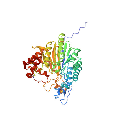
    Sphingomyelin phosphodiesterase, acid-like 3A (SMPDL3A) is a member of a small family of proteins founded by the well characterized lysosomal enzyme, acid sphingomyelinase (ASMase). ASMase converts sphingomyelin into the signaling lipid, ceramide. It was recently discovered that, in contrast to ASMase, SMPDL3A is inactive against sphingomyelin and, surprisingly, can instead hydrolyze nucleoside diphosphates and triphosphates, which may play a role in purinergic signaling. As none of the ASMase-like proteins has been structurally characterized to date, the molecular basis for their substrate preferences is unknown. Here we report crystal structures of murine SMPDL3A, which represent the first structures of an ASMase-like protein. The catalytic domain consists of a central mixed β-sandwich surrounded by α-helices. Additionally, SMPDL3A possesses a unique C-terminal domain formed from a cluster of four α-helices that appears to distinguish this protein family from other phosphoesterases. We show that SMDPL3A is a di-zinc-dependent enzyme with an active site configuration that suggests a mechanism of phosphodiester hydrolysis by a metal-activated water molecule and protonation of the leaving group by a histidine residue. Co-crystal structures of SMPDL3A with AMP and α,β-methylene ADP (AMPCP) reveal that the substrate binding site accommodates nucleotides by establishing interactions with their base, sugar, and phosphate moieties, with the latter the major contributor to binding affinity. Our study provides the structural basis for SMPDL3A substrate specificity and sheds new light on the function of ASMase-like proteins.


  • Organizational Affiliation

    From the Department of Biochemistry and Groupe de Recherche Axe sur la Structure des Proteines, Faculty of Medicine, McGill University, Montreal, Quebec H3G 0B1, Canada and.


Macromolecules
Find similar proteins by:  (by identity cutoff)  |  3D Structure
Entity ID: 1
MoleculeChains Sequence LengthOrganismDetailsImage
Acid sphingomyelinase-like phosphodiesterase 3a433Mus musculusMutation(s): 0 
Gene Names: Smpdl3aAsml3a
EC: 3.1.4
UniProt & NIH Common Fund Data Resources
Find proteins for P70158 (Mus musculus)
Explore P70158 
Go to UniProtKB:  P70158
IMPC:  MGI:1931437
Entity Groups  
Sequence Clusters30% Identity50% Identity70% Identity90% Identity95% Identity100% Identity
UniProt GroupP70158
Sequence Annotations
Expand
  • Reference Sequence
Oligosaccharides

Help

Entity ID: 2
MoleculeChains Length2D Diagram Glycosylation3D Interactions
2-acetamido-2-deoxy-beta-D-glucopyranose-(1-4)-[alpha-L-fucopyranose-(1-6)]2-acetamido-2-deoxy-beta-D-glucopyranose
B, E
3N-Glycosylation
Glycosylation Resources
GlyTouCan:  G21290RB
GlyCosmos:  G21290RB
GlyGen:  G21290RB
Entity ID: 3
MoleculeChains Length2D Diagram Glycosylation3D Interactions
2-acetamido-2-deoxy-beta-D-glucopyranose-(1-4)-2-acetamido-2-deoxy-beta-D-glucopyranose
C
2N-Glycosylation
Glycosylation Resources
GlyTouCan:  G42666HT
GlyCosmos:  G42666HT
GlyGen:  G42666HT
Entity ID: 4
MoleculeChains Length2D Diagram Glycosylation3D Interactions
alpha-D-mannopyranose-(1-3)-[alpha-D-mannopyranose-(1-6)]beta-D-mannopyranose-(1-4)-2-acetamido-2-deoxy-beta-D-glucopyranose-(1-4)-2-acetamido-2-deoxy-beta-D-glucopyranose
D
5N-Glycosylation
Glycosylation Resources
GlyTouCan:  G22768VO
GlyCosmos:  G22768VO
GlyGen:  G22768VO
Experimental Data & Validation

Experimental Data

  • Method: X-RAY DIFFRACTION
  • Resolution: 1.66 Å
  • R-Value Free: 0.191 
  • R-Value Work: 0.161 
  • R-Value Observed: 0.163 
  • Space Group: C 2 2 21
Unit Cell:
Length ( Å )Angle ( ˚ )
a = 124.351α = 90
b = 132.888β = 90
c = 80.519γ = 90
Software Package:
Software NamePurpose
PHENIXrefinement
HKL-2000data reduction
HKL-2000data scaling
PHASERphasing

Structure Validation

View Full Validation Report


Ligand Structure Quality Assessment 


Entry History 

Deposition Data

Revision History  (Full details and data files)

  • Version 1.0: 2016-01-27
    Type: Initial release
  • Version 1.1: 2016-02-03
    Changes: Database references
  • Version 1.2: 2016-03-30
    Changes: Database references
  • Version 2.0: 2020-07-29
    Type: Remediation
    Reason: Carbohydrate remediation
    Changes: Atomic model, Data collection, Database references, Derived calculations, Structure summary