5FBY | pdb_00005fby

Crystal structure of ctSPD


Experimental Data Snapshot

  • Method: X-RAY DIFFRACTION
  • Resolution: 1.90 Å
  • R-Value Free: 
    0.198 (Depositor), 0.206 (DCC) 
  • R-Value Work: 
    0.171 (Depositor), 0.182 (DCC) 
  • R-Value Observed: 
    0.172 (Depositor) 

wwPDB Validation 3D Report Full Report

Validation slider image for 5FBY

This is version 1.5 of the entry. See complete history

Literature

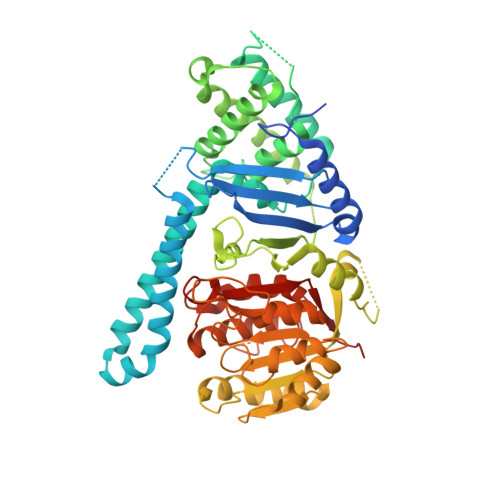
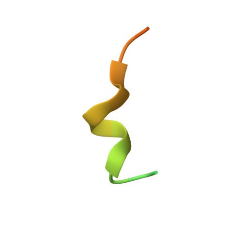
Structural basis of cohesin cleavage by separase.

Lin, Z.Luo, X.Yu, H.

(2016) Nature 532: 131-134

  • DOI: https://doi.org/10.1038/nature17402
  • Primary Citation Related Structures: 
    5FBY, 5FC2, 5FC3

  • PubMed Abstract: 

    Accurate chromosome segregation requires timely dissolution of chromosome cohesion after chromosomes are properly attached to the mitotic spindle. Separase is absolutely essential for cohesion dissolution in organisms from yeast to man. It cleaves the kleisin subunit of cohesin and opens the cohesin ring to allow chromosome segregation. Cohesin cleavage is spatiotemporally controlled by separase-associated regulatory proteins, including the inhibitory chaperone securin, and by phosphorylation of both the enzyme and substrates. Dysregulation of this process causes chromosome missegregation and aneuploidy, contributing to cancer and birth defects. Despite its essential functions, atomic structures of separase have not been determined. Here we report crystal structures of the separase protease domain from the thermophilic fungus Chaetomium thermophilum, alone or covalently bound to unphosphorylated and phosphorylated inhibitory peptides derived from a cohesin cleavage site. These structures reveal how separase recognizes cohesin and how cohesin phosphorylation by polo-like kinase 1 (Plk1) enhances cleavage. Consistent with a previous cellular study, mutating two securin residues in a conserved motif that partly matches the separase cleavage consensus converts securin from a separase inhibitor to a substrate. Our study establishes atomic mechanisms of substrate cleavage by separase and suggests competitive inhibition by securin.


  • Organizational Affiliation
    • Howard Hughes Medical Institute, University of Texas Southwestern Medical Center, 6001 Forest Park Road, Dallas, Texas 75390, USA.

Macromolecule Content 

  • Total Structure Weight: 65.34 kDa 
  • Atom Count: 4,331 
  • Modeled Residue Count: 501 
  • Deposited Residue Count: 589 
  • Unique protein chains: 2

Macromolecules

Find similar proteins by:|  3D Structure
Entity ID: 1
MoleculeChains  Sequence LengthOrganismDetailsImage
separase563Thermochaetoides thermophila DSM 1495Mutation(s): 0 
Gene Names: CTHT_0070540
EC: 3.4.22.49
UniProt
Find proteins for G0SHM3 (Chaetomium thermophilum (strain DSM 1495 / CBS 144.50 / IMI 039719))
Explore G0SHM3 
Go to UniProtKB:  G0SHM3
Entity Groups
Sequence Clusters30% Identity50% Identity70% Identity90% Identity95% Identity100% Identity
UniProt GroupG0SHM3
Sequence Annotations
Expand
Reference Sequence
Find similar proteins by:|  3D Structure
Entity ID: 2
MoleculeChains  Sequence LengthOrganismDetailsImage
cleaved peptide26synthetic constructMutation(s): 0 

Experimental Data & Validation

Experimental Data

  • Method: X-RAY DIFFRACTION
  • Resolution: 1.90 Å
  • R-Value Free:  0.198 (Depositor), 0.206 (DCC) 
  • R-Value Work:  0.171 (Depositor), 0.182 (DCC) 
  • R-Value Observed: 0.172 (Depositor) 
Space Group: P 21 21 21
Unit Cell:
Length ( Å )Angle ( ˚ )
a = 55.559α = 90
b = 98.889β = 90
c = 107.747γ = 90
Software Package:
Software NamePurpose
PHENIXrefinement
Cootmodel building
HKL-3000data collection
HKL-3000phasing

Structure Validation

View Full Validation Report


Entry History 

Deposition Data

Revision History  (Full details and data files)

  • Version 1.0: 2016-03-30
    Type: Initial release
  • Version 1.1: 2016-04-13
    Changes: Database references
  • Version 1.2: 2016-04-27
    Changes: Database references
  • Version 1.3: 2016-05-04
    Changes: Structure summary
  • Version 1.4: 2017-09-27
    Changes: Author supporting evidence, Derived calculations, Refinement description
  • Version 1.5: 2024-03-06
    Changes: Data collection, Database references