5F9K | pdb_00005f9k

Dictyostelium discoideum dUTPase at 2.2 Angstrom


Experimental Data Snapshot

  • Method: X-RAY DIFFRACTION
  • Resolution: 2.18 Å
  • R-Value Free: 
    0.251 (Depositor), 0.252 (DCC) 
  • R-Value Work: 
    0.209 (Depositor), 0.213 (DCC) 
  • R-Value Observed: 
    0.213 (Depositor) 

Starting Model: experimental
View more details

wwPDB Validation 3D Report Full Report

Validation slider image for 5F9K

Ligand Structure Quality Assessment 


This is version 1.3 of the entry. See complete history

Literature

Mitochondrial localization of Dictyostelium discoideum dUTPase mediated by its N-terminus.

Chia, C.P.Inoguchi, N.Varon, K.C.Bartholomai, B.M.Moriyama, H.

(2020) BMC Res Notes 13: 16-16

  • DOI: https://doi.org/10.1186/s13104-019-4879-7
  • Primary Citation Related Structures: 
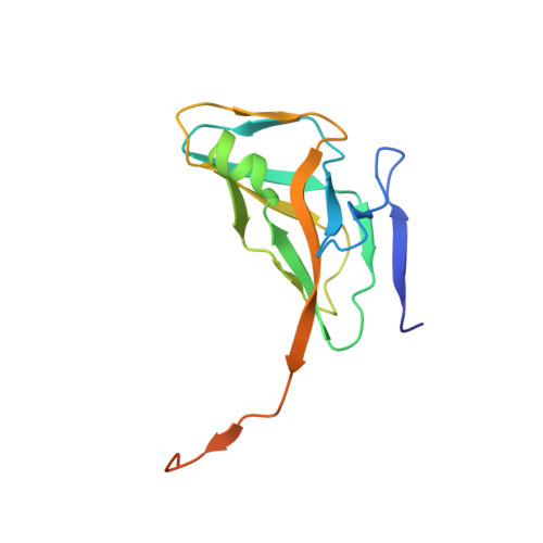
    5F9K

  • PubMed Abstract: 

    The nuclear and mitochondrial genomes of Dictyostelium discoideum, a unicellular eukaryote, have relatively high A+T-contents of 77.5% and 72.65%, respectively. To begin to investigate how the pyrimidine biosynthetic pathway fulfills the demand for dTTP, we determined the catalytic properties and structure of the key enzyme deoxyuridine triphosphate nucleotidohydrolase (dUTPase) that hydrolyzes dUTP to dUMP, the precursor of dTTP. The annotated genome of D. discoideum identifies a gene encoding a polypeptide containing the five conserved motifs of homotrimeric dUTPases. Recombinant proteins, comprised of either full-length or core polypeptides with all conserved motifs but lacking residues 1-37 of the N-terminus, were active dUTPases. Crystallographic analyses of the core enzyme indicated that the C-termini, normally flexible, were constrained by interactions with the shortened N-termini that arose from the loss of residues 1-37. This allowed greater access of dUTP to active sites, resulting in enhanced catalytic parameters. A tagged protein comprised of the N-terminal forty amino acids of dUTPase fused to green fluorescent protein (GFP) was expressed in D. discoideum cells. Supporting a prediction of mitochondrial targeting information within the N-terminus, localization and subcellular fractionation studies showed GFP to be in mitochondria. N-terminal sequencing of immunoprecipitated GFP revealed the loss of the dUTPase sequence upon import into the organelle.


  • Organizational Affiliation
    • School of Biological Sciences, Univ. Nebraska-Lincoln, Lincoln, NE, 68588-0118, USA. cchia1@unl.edu.

Macromolecule Content 

  • Total Structure Weight: 48.24 kDa 
  • Atom Count: 3,181 
  • Modeled Residue Count: 386 
  • Deposited Residue Count: 438 
  • Unique protein chains: 1

Macromolecules

Find similar proteins by:|  3D Structure
Entity ID: 1
MoleculeChains  Sequence LengthOrganismDetailsImage
Deoxyuridine 5'-triphosphate nucleotidohydrolase
A, B, C
146Dictyostelium discoideumMutation(s): 0 
Gene Names: dutDDB_G0293374
EC: 3.6.1.23
UniProt
Find proteins for Q54BW5 (Dictyostelium discoideum)
Explore Q54BW5 
Go to UniProtKB:  Q54BW5
Entity Groups
Sequence Clusters30% Identity50% Identity70% Identity90% Identity95% Identity100% Identity
UniProt GroupQ54BW5
Sequence Annotations
Expand
Reference Sequence

Small Molecules

Ligands 3 Unique
IDChains Name / Formula / InChI Key2D Diagram3D Interactions
DUP

Query on DUP


Download:Ideal Coordinates CCD File
D [auth A],
E [auth B],
L [auth C]
2'-DEOXYURIDINE 5'-ALPHA,BETA-IMIDO-TRIPHOSPHATE
C9 H16 N3 O13 P3
XZLLMTSKYYYJLH-SHYZEUOFSA-N
SO4

Query on SO4


Download:Ideal Coordinates CCD File
K [auth C]SULFATE ION
O4 S
QAOWNCQODCNURD-UHFFFAOYSA-L
MG

Query on MG


Download:Ideal Coordinates CCD File
F [auth B],
G [auth B],
H [auth B],
I [auth B],
J [auth C]
MAGNESIUM ION
Mg
JLVVSXFLKOJNIY-UHFFFAOYSA-N

Experimental Data & Validation

Experimental Data

  • Method: X-RAY DIFFRACTION
  • Resolution: 2.18 Å
  • R-Value Free:  0.251 (Depositor), 0.252 (DCC) 
  • R-Value Work:  0.209 (Depositor), 0.213 (DCC) 
  • R-Value Observed: 0.213 (Depositor) 
Space Group: P 21 21 21
Unit Cell:
Length ( Å )Angle ( ˚ )
a = 71.633α = 90
b = 72.458β = 90
c = 75.158γ = 90
Software Package:
Software NamePurpose
PHENIXrefinement
HKL-2000data reduction
HKL-2000data scaling
PHENIXphasing

Structure Validation

View Full Validation Report


Ligand Structure Quality Assessment 


Entry History 

& Funding Information

Deposition Data


Funding OrganizationLocationGrant Number
The John C. and Nettie V. David Memorial Trust Fund of the University of Nebraska FoundationUnited States--

Revision History  (Full details and data files)

  • Version 1.0: 2016-12-14
    Type: Initial release
  • Version 1.1: 2017-09-27
    Changes: Author supporting evidence
  • Version 1.2: 2020-01-22
    Changes: Database references
  • Version 1.3: 2023-09-27
    Changes: Data collection, Database references, Derived calculations, Refinement description