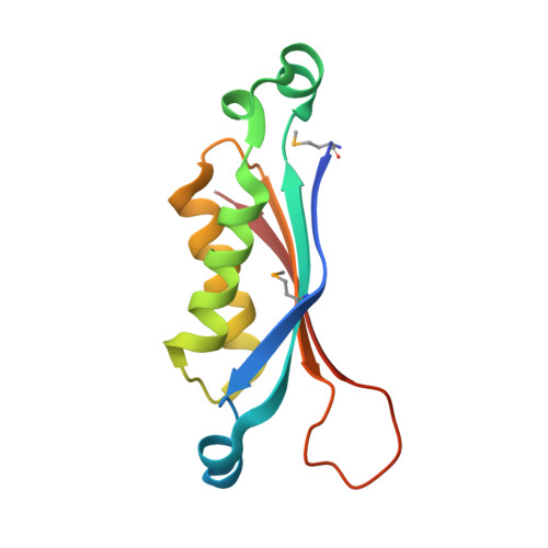
Crystal structure of dihydroneopterin aldolase from Bacillus anthracis complexed with L-neopterin at 1.5 Angstroms resolution .
Maltseva, N., Kim, Y., Shatsman, S., Anderson, W.F., Joachimiak, A., CSGID, Center for Structural Genomics of Infectious Diseases (CSGID)To be published.