5F2A

Crystal structure of murine neuroglobin mutant F106W at 310 MPa pressure


Experimental Data Snapshot

  • Method: X-RAY DIFFRACTION
  • Resolution: 2.10 Å
  • R-Value Free: 0.260 
  • R-Value Work: 0.165 
  • R-Value Observed: 0.169 

wwPDB Validation   3D Report Full Report


Ligand Structure Quality Assessment 


This is version 1.3 of the entry. See complete history


Literature

Determinants of neuroglobin plasticity highlighted by joint coarse-grained simulations and high pressure crystallography.

Colloc'h, N.Sacquin-Mora, S.Avella, G.Dhaussy, A.C.Prange, T.Vallone, B.Girard, E.

(2017) Sci Rep 7: 1858-1858

  • DOI: https://doi.org/10.1038/s41598-017-02097-1
  • Primary Citation of Related Structures:  
    5EET, 5EOH, 5EQM, 5EU2, 5EV5, 5EYJ, 5EYS, 5F0B, 5F2A

  • PubMed Abstract: 

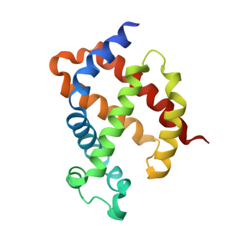
    Investigating the effect of pressure sheds light on the dynamics and plasticity of proteins, intrinsically correlated to functional efficiency. Here we detail the structural response to pressure of neuroglobin (Ngb), a hexacoordinate globin likely to be involved in neuroprotection. In murine Ngb, reversible coordination is achieved by repositioning the heme more deeply into a large internal cavity, the "heme sliding mechanism". Combining high pressure crystallography and coarse-grain simulations on wild type Ngb as well as two mutants, one (V101F) with unaffected and another (F106W) with decreased affinity for CO, we show that Ngb hinges around a rigid mechanical nucleus of five hydrophobic residues (V68, I72, V109, L113, Y137) during its conformational transition induced by gaseous ligand, that the intrinsic flexibility of the F-G loop appears essential to drive the heme sliding mechanism, and that residue Val 101 may act as a sensor of the interaction disruption between the heme and the distal histidine.


  • Organizational Affiliation

    ISTCT CNRS UNICAEN CEA Normandie Univ., CERVOxy team, centre Cyceron, 14000, Caen, France. colloch@cyceron.fr.


Macromolecules
Find similar proteins by:  (by identity cutoff)  |  3D Structure
Entity ID: 1
MoleculeChains Sequence LengthOrganismDetailsImage
Neuroglobin154Mus musculusMutation(s): 3 
Gene Names: Ngb
UniProt
Find proteins for Q9ER97 (Mus musculus)
Explore Q9ER97 
Go to UniProtKB:  Q9ER97
Entity Groups  
Sequence Clusters30% Identity50% Identity70% Identity90% Identity95% Identity100% Identity
UniProt GroupQ9ER97
Sequence Annotations
Expand
  • Reference Sequence
Small Molecules
Ligands 2 Unique
IDChains Name / Formula / InChI Key2D Diagram3D Interactions
HEM
Query on HEM

Download Ideal Coordinates CCD File 
B [auth A]PROTOPORPHYRIN IX CONTAINING FE
C34 H32 Fe N4 O4
KABFMIBPWCXCRK-RGGAHWMASA-L
SO4
Query on SO4

Download Ideal Coordinates CCD File 
C [auth A]SULFATE ION
O4 S
QAOWNCQODCNURD-UHFFFAOYSA-L
Experimental Data & Validation

Experimental Data

  • Method: X-RAY DIFFRACTION
  • Resolution: 2.10 Å
  • R-Value Free: 0.260 
  • R-Value Work: 0.165 
  • R-Value Observed: 0.169 
  • Space Group: H 3 2
Unit Cell:
Length ( Å )Angle ( ˚ )
a = 85.99α = 90
b = 85.99β = 90
c = 114.578γ = 120
Software Package:
Software NamePurpose
SCALAdata scaling
PHASERphasing
DMphasing
REFMACrefinement
PDB_EXTRACTdata extraction
XDSdata reduction

Structure Validation

View Full Validation Report


Ligand Structure Quality Assessment 


Entry History 

Deposition Data

Revision History  (Full details and data files)

  • Version 1.0: 2016-10-19
    Type: Initial release
  • Version 1.1: 2017-05-31
    Changes: Database references
  • Version 1.2: 2021-07-28
    Changes: Structure summary
  • Version 1.3: 2024-01-10
    Changes: Data collection, Database references, Refinement description