5EIZ | pdb_00005eiz

Crystal structure of Y99A mutant of human macrophage migration inhibitory factor


Experimental Data Snapshot

  • Method: X-RAY DIFFRACTION
  • Resolution: 1.96 Å
  • R-Value Free: 
    0.181 (Depositor), 0.190 (DCC) 
  • R-Value Work: 
    0.157 (Depositor), 0.170 (DCC) 
  • R-Value Observed: 
    0.158 (Depositor) 

Starting Model: experimental
View more details

wwPDB Validation 3D Report Full Report

Validation slider image for 5EIZ

This is version 1.2 of the entry. See complete history

Literature

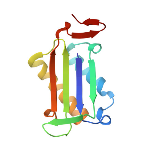
Nanosecond Dynamics Regulate the MIF-Induced Activity of CD74.

Pantouris, G.Ho, J.Shah, D.Syed, M.A.Leng, L.Bhandari, V.Bucala, R.Batista, V.S.Loria, J.P.Lolis, E.J.

(2018) Angew Chem Int Ed Engl 

  • DOI: https://doi.org/10.1002/anie.201803191
  • Primary Citation Related Structures: 
    5EIZ, 5UZY, 6BG6, 6BG7

  • PubMed Abstract: 

    Macrophage migration inhibitory factor (MIF) activates CD74, which leads to severe disorders including inflammation, autoimmune diseases and cancer under pathological conditions. Molecular dynamics (MD) simulations up to one microsecond revealed dynamical correlation between a residue located at the opening of one end of the MIF solvent channel, previously thought to be a consequence of homotrimerization, and residues in a distal region responsible for CD74 activation. Experiments verified the allosteric regulatory site and identified a pathway to this site via the MIF β-strands. The reported findings provide fundamental insights on a dynamic mechanism that controls the MIF-induced activation of CD74.


  • Organizational Affiliation
    • Department of Pharmacology, School of Medicine, Yale University, New Haven, CT, 06510, USA.

Macromolecule Content 

  • Total Structure Weight: 37.47 kDa 
  • Atom Count: 2,888 
  • Modeled Residue Count: 342 
  • Deposited Residue Count: 342 
  • Unique protein chains: 1

Macromolecules

Find similar proteins by:|  3D Structure
Entity ID: 1
MoleculeChains  Sequence LengthOrganismDetailsImage
Macrophage migration inhibitory factor
A, B, C
114Homo sapiensMutation(s): 1 
Gene Names: MIFGLIFMMIF
EC: 5.3.2.1 (PDB Primary Data), 5.3.3.12 (PDB Primary Data)
UniProt & NIH Common Fund Data Resources
Find proteins for P14174 (Homo sapiens)
Explore P14174 
Go to UniProtKB:  P14174
PHAROS:  P14174
GTEx:  ENSG00000240972 
Entity Groups
Sequence Clusters30% Identity50% Identity70% Identity90% Identity95% Identity100% Identity
UniProt GroupP14174
Sequence Annotations
Expand
Reference Sequence

Small Molecules

Ligands 3 Unique
IDChains Name / Formula / InChI Key2D Diagram3D Interactions
SO4

Query on SO4


Download:Ideal Coordinates CCD File
E [auth A],
H [auth B],
K [auth C]
SULFATE ION
O4 S
QAOWNCQODCNURD-UHFFFAOYSA-L
GOL

Query on GOL


Download:Ideal Coordinates CCD File
D [auth A],
G [auth B],
J [auth C]
GLYCEROL
C3 H8 O3
PEDCQBHIVMGVHV-UHFFFAOYSA-N
IPA

Query on IPA


Download:Ideal Coordinates CCD File
F [auth A],
I [auth B]
ISOPROPYL ALCOHOL
C3 H8 O
KFZMGEQAYNKOFK-UHFFFAOYSA-N

Experimental Data & Validation

Experimental Data

  • Method: X-RAY DIFFRACTION
  • Resolution: 1.96 Å
  • R-Value Free:  0.181 (Depositor), 0.190 (DCC) 
  • R-Value Work:  0.157 (Depositor), 0.170 (DCC) 
  • R-Value Observed: 0.158 (Depositor) 
Space Group: P 21 21 21
Unit Cell:
Length ( Å )Angle ( ˚ )
a = 67.696α = 90
b = 67.958β = 90
c = 86.519γ = 90
Software Package:
Software NamePurpose
HKL-2000data collection
HKL-2000data scaling
REFMACrefinement
PDB_EXTRACTdata extraction
DENZOdata reduction
PHASERphasing

Structure Validation

View Full Validation Report


Entry History 

Deposition Data

Revision History  (Full details and data files)

  • Version 1.0: 2015-11-11
    Type: Initial release
  • Version 1.1: 2018-06-06
    Changes: Data collection, Database references, Derived calculations
  • Version 1.2: 2023-09-27
    Changes: Data collection, Database references, Refinement description