5EI3

Co-crystal structure of eIF4E with nucleotide mimetic inhibitor.


Experimental Data Snapshot

  • Method: X-RAY DIFFRACTION
  • Resolution: 1.71 Å
  • R-Value Free: 0.187 
  • R-Value Work: 0.166 
  • R-Value Observed: 0.167 

wwPDB Validation   3D Report Full Report


Ligand Structure Quality Assessment 


This is version 2.0 of the entry. See complete history


Literature

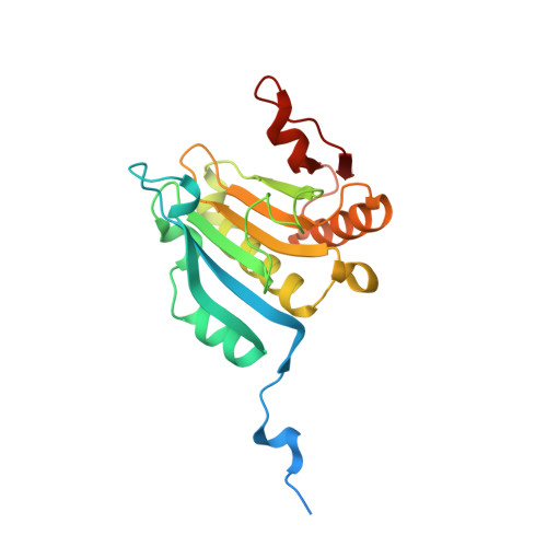
Design of nucleotide-mimetic and non-nucleotide inhibitors of the translation initiation factor eIF4E: Synthesis, structural and functional characterisation.

Soukarieh, F.Nowicki, M.W.Bastide, A.Poyry, T.Jones, C.Dudek, K.Patwardhan, G.Meullenet, F.Oldham, N.J.Walkinshaw, M.D.Willis, A.E.Fischer, P.M.

(2016) Eur J Med Chem 124: 200-217

  • DOI: https://doi.org/10.1016/j.ejmech.2016.08.047
  • Primary Citation of Related Structures:  
    5EHC, 5EI3, 5EIR, 5EKV

  • PubMed Abstract: 

    Eukaryotic translation initiation factor 4E (eIF4E) is considered as the corner stone in the cap-dependent translation initiation machinery. Its role is to recruit mRNA to the ribosome through recognition of the 5'-terminal mRNA cap structure (m 7 GpppN, where G is guanosine, N is any nucleotide). eIF4E is implicated in cell transformation, tumourigenesis, and angiogenesis by facilitating translation of oncogenic mRNAs; it is thus regarded as an attractive anticancer drug target. We have used two approaches to design cap-binding inhibitors of eIF4E by modifying the N 7 -substituent of m 7 GMP and replacing the phosphate group with isosteres such as squaramides, sulfonamides, and tetrazoles, as well as by structure-based virtual screening aimed at identifying non-nucleotide cap-binding antagonists. Phosphomimetic nucleotide derivatives and highly ranking virtual hits were evaluated in a series of in vitro and cell-based assays to identify the first non-nucleotide eIF4E cap-binding inhibitor with activities in cell-based assays, N-[(5,6-dihydro-6-oxo-1,3-dioxolo[4,5-g]quinolin-7-yl)methyl]-N'-(2-methyl-propyl)-N-(phenyl-methyl)thiourea (14), including down-regulation of oncogenic proteins and suppression of RNA incorporation into polysomes. Although we did not observe cellular activity with any of our modified m 7 GMP phosphate isostere compounds, we obtained X-ray crystallography structures of three such compounds in complex with eIF4E, 5'-deoxy-5'-(1,2-dioxo-3-hydroxycyclobut-3-en-4-yl)amino-N 7 -methyl-guanosine (4a), N 7 -3-chlorobenzyl-5'-deoxy-5'-(1,2-dioxo-3-hydroxy-cyclobut-3-en-4-yl)amino-guanosine (4f), and N 7 -benzyl-5'-deoxy-5'-(trifluoromethyl-sulfamoyl)guanosine (7a). Collectively, the data we present on structure-based design of eIF4E cap-binding inhibitors should facilitate the optimisation of such compounds as potential anticancer agents.


  • Organizational Affiliation

    School of Pharmacy and Centre for Biomolecular Sciences, University of Nottingham, University Park, Nottingham NG7 2RD, UK.


Macromolecules
Find similar proteins by:  (by identity cutoff)  |  3D Structure
Entity ID: 1
MoleculeChains Sequence LengthOrganismDetailsImage
Eukaryotic translation initiation factor 4E217Homo sapiensMutation(s): 0 
Gene Names: EIF4EEIF4EL1EIF4F
UniProt & NIH Common Fund Data Resources
Find proteins for P06730 (Homo sapiens)
Explore P06730 
Go to UniProtKB:  P06730
PHAROS:  P06730
GTEx:  ENSG00000151247 
Entity Groups  
Sequence Clusters30% Identity50% Identity70% Identity90% Identity95% Identity100% Identity
UniProt GroupP06730
Sequence Annotations
Expand
  • Reference Sequence

Find similar proteins by:  Sequence   |   3D Structure  

Entity ID: 2
MoleculeChains Sequence LengthOrganismDetailsImage
Eukaryotic translation initiation factor 4 gamma14synthetic constructMutation(s): 0 
UniProt & NIH Common Fund Data Resources
Find proteins for Q04637 (Homo sapiens)
Explore Q04637 
Go to UniProtKB:  Q04637
PHAROS:  Q04637
GTEx:  ENSG00000114867 
Entity Groups  
Sequence Clusters30% Identity50% Identity70% Identity90% Identity95% Identity100% Identity
UniProt GroupQ04637
Sequence Annotations
Expand
  • Reference Sequence
Small Molecules
Ligands 2 Unique
IDChains Name / Formula / InChI Key2D Diagram3D Interactions
5O8
Query on 5O8

Download Ideal Coordinates CCD File 
C [auth A]~{N}-[[(2~{R},3~{S},4~{R},5~{R})-5-[2-azanyl-6-oxidanylidene-7-(phenylmethyl)-1~{H}-purin-7-ium-9-yl]-3,4-bis(oxidanyl)oxolan-2-yl]methyl]-1,1,1-tris(fluoranyl)methanesulfonamide
C18 H20 F3 N6 O6 S
NMFHLBUCTTXCHX-XNIJJKJLSA-O
SO4
Query on SO4

Download Ideal Coordinates CCD File 
D [auth A]SULFATE ION
O4 S
QAOWNCQODCNURD-UHFFFAOYSA-L
Binding Affinity Annotations 
IDSourceBinding Affinity
5O8 Binding MOAD:  5EI3 Kd: 5.65e+4 (nM) from 1 assay(s)
Experimental Data & Validation

Experimental Data

  • Method: X-RAY DIFFRACTION
  • Resolution: 1.71 Å
  • R-Value Free: 0.187 
  • R-Value Work: 0.166 
  • R-Value Observed: 0.167 
  • Space Group: P 21 2 21
Unit Cell:
Length ( Å )Angle ( ˚ )
a = 38.59α = 90
b = 52.08β = 90
c = 125.19γ = 90
Software Package:
Software NamePurpose
SCALAdata scaling
PHENIXrefinement
PDB_EXTRACTdata extraction
MOSFLMdata reduction
PHENIXphasing

Structure Validation

View Full Validation Report


Ligand Structure Quality Assessment 


Entry History 

Deposition Data

Revision History  (Full details and data files)

  • Version 1.0: 2016-09-07
    Type: Initial release
  • Version 1.1: 2016-09-14
    Changes: Database references
  • Version 1.2: 2017-11-29
    Changes: Database references
  • Version 2.0: 2024-01-10
    Changes: Atomic model, Data collection, Database references, Refinement description