


X-ray structure uridine phosphorylase from Vibrio cholerae in complex with uridine at 2.24 A resolution
Prokofev, I.I., Lashkov, A.A., Gabdoulkhakov, A.G., Betzel, C., Mikhailov, A.M.To be published.
Experimental Data Snapshot
Starting Model: experimental
View more details
Entity ID: 1 | |||||
|---|---|---|---|---|---|
| Molecule | Chains | Sequence Length | Organism | Details | Image |
| Uridine phosphorylase | 253 | Vibrio cholerae | Mutation(s): 0 Gene Names: udp, udp_1, DN30_1909, EN12_05055, ERS013138_02408, ERS013140_00580, ERS013186_00327, ERS013199_00063, ERS013201_00032, ERS013202_00369... EC: 2.4.2.3 | 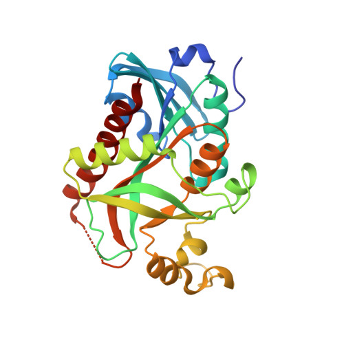 | |
UniProt | |||||
Entity Groups | |||||
| Sequence Clusters | 30% Identity50% Identity70% Identity90% Identity95% Identity100% Identity | ||||
| UniProt Group | Q9K4U1 | ||||
Sequence AnnotationsExpand | |||||
Reference Sequence | |||||
| Ligands 7 Unique | |||||
|---|---|---|---|---|---|
| ID | Chains | Name / Formula / InChI Key | 2D Diagram | 3D Interactions | |
| CTN Download:Ideal Coordinates CCD File | W [auth E], Z [auth F] | 4-AMINO-1-BETA-D-RIBOFURANOSYL-2(1H)-PYRIMIDINONE C9 H13 N3 O5 UHDGCWIWMRVCDJ-XVFCMESISA-N |  | ||
| TRS Download:Ideal Coordinates CCD File | I [auth A] | 2-AMINO-2-HYDROXYMETHYL-PROPANE-1,3-DIOL C4 H12 N O3 LENZDBCJOHFCAS-UHFFFAOYSA-O |  | ||
| CYT Download:Ideal Coordinates CCD File | H [auth A] K [auth B] N [auth C] O [auth C] R [auth D] | 6-AMINOPYRIMIDIN-2(1H)-ONE C4 H5 N3 O OPTASPLRGRRNAP-UHFFFAOYSA-N |  | ||
| PEG Download:Ideal Coordinates CCD File | M [auth C] | DI(HYDROXYETHYL)ETHER C4 H10 O3 MTHSVFCYNBDYFN-UHFFFAOYSA-N |  | ||
| GOL Download:Ideal Coordinates CCD File | J [auth B], L [auth C], T [auth D], V [auth E] | GLYCEROL C3 H8 O3 PEDCQBHIVMGVHV-UHFFFAOYSA-N |  | ||
| EDO Download:Ideal Coordinates CCD File | Q [auth D], X [auth F], Y [auth F] | 1,2-ETHANEDIOL C2 H6 O2 LYCAIKOWRPUZTN-UHFFFAOYSA-N |  | ||
| NA Download:Ideal Coordinates CCD File | G [auth A], P [auth C], U [auth E] | SODIUM ION Na FKNQFGJONOIPTF-UHFFFAOYSA-N |  | ||
| Length ( Å ) | Angle ( ˚ ) |
|---|---|
| a = 59.949 | α = 67.54 |
| b = 76.336 | β = 73.74 |
| c = 89.694 | γ = 84.92 |
| Software Name | Purpose |
|---|---|
| SCALA | data scaling |
| MOLREP | phasing |
| REFMAC | refinement |
| PDB_EXTRACT | data extraction |
| XDS | data reduction |
| Funding Organization | Location | Grant Number |
|---|---|---|
| RFBR | Russian Federation | 14-04-00952a |


