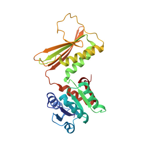
5EES

Crystal structure of DapB in complex with NADP+ from Corynebacterium glutamicum


Experimental Data Snapshot

  • Method: X-RAY DIFFRACTION
  • Resolution: 2.15 Å
  • R-Value Free: 0.211 
  • R-Value Work: 0.179 
  • R-Value Observed: 0.180 

Starting Model: experimental
View more details

wwPDB Validation   3D Report Full Report


Ligand Structure Quality Assessment 


This is version 1.3 of the entry. See complete history


Literature

Structural Insight into Dihydrodipicolinate Reductase from Corybebacterium glutamicum for Lysine Biosynthesis.

Sagong, H.Y.Kim, K.J.

(2016) J Microbiol Biotechnol 26: 226-232

  • DOI: https://doi.org/10.4014/jmb.1508.08086
  • Primary Citation of Related Structures:  
    5EER, 5EES

  • PubMed Abstract: 

    Dihydrodipicolinate reductase is an enzyme that converts dihydrodipicolinate to tetrahydrodipicolinate using an NAD(P)H cofactor in L-lysine biosynthesis. To increase the understanding of the molecular mechanisms of lysine biosynthesis, we determined the crystal structure of dihydrodipicolinate reductase from Corynebacterium glutamicum (CgDapB). CgDapB functions as a tetramer, and each protomer is composed of two domains, an Nterminal domain and a C-terminal domain. The N-terminal domain mainly contributes to nucleotide binding, whereas the C-terminal domain is involved in substrate binding. We elucidated the mode of cofactor binding to CgDapB by determining the crystal structure of the enzyme in complex with NADP(+) and found that CgDapB utilizes both NADH and NADPH as cofactors. Moreover, we determined the substrate binding mode of the enzyme based on the coordination mode of two sulfate ions in our structure. Compared with Mycobacterium tuberculosis DapB in complex with its cofactor and inhibitor, we propose that the domain movement for active site constitution occurs when both cofactor and substrate bind to the enzyme.


  • Organizational Affiliation

    School of Life Sciences, KNU Creative BioResearch Group, Kyungpook National University, Daegu 41566, Republic of Korea.


Macromolecules
Find similar proteins by:  (by identity cutoff)  |  3D Structure
Entity ID: 1
MoleculeChains Sequence LengthOrganismDetailsImage
4-hydroxy-tetrahydrodipicolinate reductase247Corynebacterium glutamicum ATCC 13032Mutation(s): 0 
Gene Names: dapBCgl1973cg2163
EC: 1.17.1.8
UniProt
Find proteins for P40110 (Corynebacterium glutamicum (strain ATCC 13032 / DSM 20300 / JCM 1318 / BCRC 11384 / CCUG 27702 / LMG 3730 / NBRC 12168 / NCIMB 10025 / NRRL B-2784 / 534))
Explore P40110 
Go to UniProtKB:  P40110
Entity Groups  
Sequence Clusters30% Identity50% Identity70% Identity90% Identity95% Identity100% Identity
UniProt GroupP40110
Sequence Annotations
Expand
  • Reference Sequence
Experimental Data & Validation

Experimental Data

  • Method: X-RAY DIFFRACTION
  • Resolution: 2.15 Å
  • R-Value Free: 0.211 
  • R-Value Work: 0.179 
  • R-Value Observed: 0.180 
  • Space Group: I 41 2 2
Unit Cell:
Length ( Å )Angle ( ˚ )
a = 107.969α = 90
b = 107.969β = 90
c = 172.049γ = 90
Software Package:
Software NamePurpose
HKL-2000data collection
REFMACrefinement
PDB_EXTRACTdata extraction
HKL-2000data reduction
HKL-2000data scaling
CNSphasing

Structure Validation

View Full Validation Report


Ligand Structure Quality Assessment 


Entry History 

Deposition Data

Revision History  (Full details and data files)

  • Version 1.0: 2015-11-11
    Type: Initial release
  • Version 1.1: 2017-07-19
    Changes: Database references, Derived calculations
  • Version 1.2: 2020-09-09
    Changes: Structure summary
  • Version 1.3: 2023-11-08
    Changes: Data collection, Database references, Refinement description