5E66 | pdb_00005e66

The complex structure of Hemagglutinin-esterase-fusion mutant protein from the influenza D virus with receptor analog 9-N-Ac-Sia


Experimental Data Snapshot

  • Method: X-RAY DIFFRACTION
  • Resolution: 3.10 Å
  • R-Value Free: 
    0.265 (Depositor), 0.270 (DCC) 
  • R-Value Work: 
    0.234 (Depositor), 0.238 (DCC) 
  • R-Value Observed: 
    0.235 (Depositor) 

Starting Model: experimental
View more details

wwPDB Validation 3D Report Full Report

Validation slider image for 5E66

Ligand Structure Quality Assessment 


This is version 2.2 of the entry. See complete history

Literature

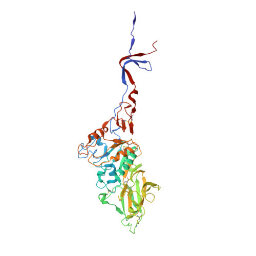
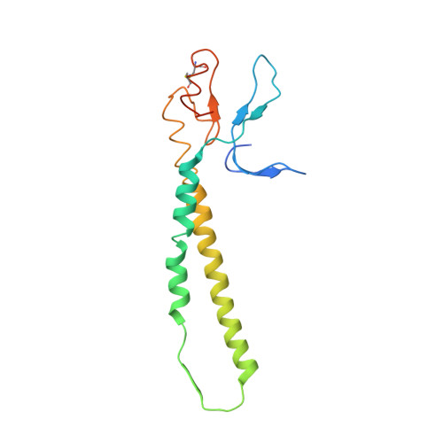
An Open Receptor-Binding Cavity of Hemagglutinin-Esterase-Fusion Glycoprotein from Newly-Identified Influenza D Virus: Basis for Its Broad Cell Tropism

Song, H.Qi, J.Khedri, Z.Diaz, S.Yu, H.Chen, X.Varki, A.Shi, Y.Gao, G.F.

(2016) PLoS Pathog 12: e1005411-e1005411

  • DOI: https://doi.org/10.1371/journal.ppat.1005411
  • Primary Citation Related Structures: 
    5E5W, 5E62, 5E64, 5E65, 5E66

  • PubMed Abstract: 

    Influenza viruses cause seasonal flu each year and pandemics or epidemic sporadically, posing a major threat to public health. Recently, a new influenza D virus (IDV) was isolated from pigs and cattle. Here, we reveal that the IDV utilizes 9-O-acetylated sialic acids as its receptor for virus entry. Then, we determined the crystal structures of hemagglutinin-esterase-fusion glycoprotein (HEF) of IDV both in its free form and in complex with the receptor and enzymatic substrate analogs. The IDV HEF shows an extremely similar structural fold as the human-infecting influenza C virus (ICV) HEF. However, IDV HEF has an open receptor-binding cavity to accommodate diverse extended glycan moieties. This structural difference provides an explanation for the phenomenon that the IDV has a broad cell tropism. As IDV HEF is structurally and functionally similar to ICV HEF, our findings highlight the potential threat of the virus to public health.


  • Organizational Affiliation
    • CAS Key Laboratory of Pathogenic Microbiology and Immunology, Institute of Microbiology, Chinese Academy of Sciences, Beijing, China.

Macromolecule Content 

  • Total Structure Weight: 67.5 kDa 
  • Atom Count: 4,575 
  • Modeled Residue Count: 573 
  • Deposited Residue Count: 593 
  • Unique protein chains: 2

Macromolecules

Find similar proteins by:|  3D Structure
Entity ID: 1
MoleculeChains  Sequence LengthOrganismDetailsImage
Hemagglutinin-esterase427Influenza D virus (D/swine/Oklahoma/1334/2011)Mutation(s): 0 
Gene Names: HEF
EC: 3.1.1.53
UniProt
Find proteins for K9LG83 (Influenza D virus)
Explore K9LG83 
Go to UniProtKB:  K9LG83
Entity Groups
Sequence Clusters30% Identity50% Identity70% Identity90% Identity95% Identity100% Identity
UniProt GroupK9LG83
Glycosylation
Glycosylation Sites: 3
Sequence Annotations
Expand
Reference Sequence
Find similar proteins by:|  3D Structure
Entity ID: 2
MoleculeChains  Sequence LengthOrganismDetailsImage
Hemagglutinin-esterase166Influenza D virus (D/swine/Oklahoma/1334/2011)Mutation(s): 0 
Gene Names: HEF
EC: 3.1.1.53
UniProt
Find proteins for K9LG83 (Influenza D virus)
Explore K9LG83 
Go to UniProtKB:  K9LG83
Entity Groups
Sequence Clusters30% Identity50% Identity70% Identity90% Identity95% Identity100% Identity
UniProt GroupK9LG83
Glycosylation
Glycosylation Sites: 2
Sequence Annotations
Expand
Reference Sequence

Oligosaccharides

Help  
Entity ID: 3
MoleculeChains Length2D Diagram GlycosylationD Interactions
alpha-D-mannopyranose-(1-3)-[alpha-D-mannopyranose-(1-6)]alpha-D-mannopyranose-(1-6)-beta-D-mannopyranose-(1-4)-2-acetamido-2-deoxy-beta-D-glucopyranose-(1-4)-2-acetamido-2-deoxy-beta-D-glucopyranose
C
6N-Glycosylation
Glycosylation Resources
GlyTouCan: G94106MV
GlyCosmos: G94106MV
GlyGen: G94106MV
Entity ID: 4
MoleculeChains Length2D Diagram GlycosylationD Interactions
2-acetamido-2-deoxy-beta-D-glucopyranose-(1-4)-2-acetamido-2-deoxy-beta-D-glucopyranose
D, E, F
2N-Glycosylation
Glycosylation Resources
GlyTouCan: G42666HT
GlyCosmos: G42666HT
GlyGen: G42666HT

Experimental Data & Validation

Experimental Data

  • Method: X-RAY DIFFRACTION
  • Resolution: 3.10 Å
  • R-Value Free:  0.265 (Depositor), 0.270 (DCC) 
  • R-Value Work:  0.234 (Depositor), 0.238 (DCC) 
  • R-Value Observed: 0.235 (Depositor) 
Space Group: I 21 3
Unit Cell:
Length ( Å )Angle ( ˚ )
a = 165.297α = 90
b = 165.297β = 90
c = 165.297γ = 90
Software Package:
Software NamePurpose
PHENIXrefinement
PHASERphasing
HKL-2000data reduction
HKL-2000data scaling

Structure Validation

View Full Validation Report


Ligand Structure Quality Assessment 


Entry History 

Deposition Data

Revision History  (Full details and data files)

  • Version 1.0: 2016-03-23
    Type: Initial release
  • Version 1.1: 2018-09-05
    Changes: Data collection, Database references, Derived calculations
  • Version 2.0: 2020-07-29
    Type: Remediation
    Reason: Carbohydrate remediation
    Changes: Advisory, Atomic model, Data collection, Derived calculations, Structure summary
  • Version 2.1: 2023-11-08
    Changes: Data collection, Database references, Derived calculations, Refinement description, Structure summary
  • Version 2.2: 2024-11-06
    Changes: Structure summary