5E4K

Structure of ligand binding region of uPARAP at pH 7.4


Experimental Data Snapshot

  • Method: X-RAY DIFFRACTION
  • Resolution: 2.58 Å
  • R-Value Free: 0.257 
  • R-Value Work: 0.214 
  • R-Value Observed: 0.216 

wwPDB Validation   3D Report Full Report


Ligand Structure Quality Assessment 


This is version 1.2 of the entry. See complete history


Literature

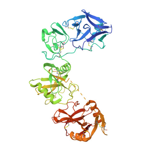
Crystal structures of the ligand-binding region of uPARAP: effect of calcium ion binding

Yuan, C.Jurgensen, H.J.Engelholm, L.H.Li, R.Liu, M.Jiang, L.Luo, Z.Behrendt, N.Huang, M.

(2016) Biochem J 473: 2359-2368

  • DOI: https://doi.org/10.1042/BCJ20160276
  • Primary Citation of Related Structures:  
    5E4K, 5EW6

  • PubMed Abstract: 

    The proteins of the mannose receptor (MR) family share a common domain organization and have a broad range of biological functions. Urokinase plasminogen activator receptor-associated protein (uPARAP) (or Endo180) is a member of this family and plays an important role in extracellular matrix remodelling through interaction with its ligands, including collagens and urokinase plasminogen activator receptor (uPAR). We report the crystal structures of the first four domains of uPARAP (also named the ligand-binding region, LBR) at pH 7.4 in Ca(2+)-bound and Ca(2+)-free forms. The first domain (cysteine-rich or CysR domain) folds into a new and unique conformation different from the β-trefoil fold of typical CysR domains. The so-called long loop regions (LLRs) of the C-type lectin-like domain (CTLD) 1 and 2 (the third and fourth domain) mediate the direct contacts between these domains. These LLRs undergo a Ca(2+)-dependent conformational change, and this is likely to be the key structural determinant affecting the overall conformation of uPARAP. Our results provide a molecular mechanism to support the structural flexibility of uPARAP, and shed light on the structural flexibility of other members of the MR family.


  • Organizational Affiliation

    State Key Laboratory of Structural Chemistry, Fujian Institute of Research on the Structure of Matter, Chinese Academy of Sciences, Fuzhou 350002, China College of Bioscience and Biotechnology, Fuzhou University, Fuzhou 350108, China.


Macromolecules
Find similar proteins by:  (by identity cutoff)  |  3D Structure
Entity ID: 1
MoleculeChains Sequence LengthOrganismDetailsImage
C-type mannose receptor 2492Homo sapiensMutation(s): 1 
Gene Names: MRC2CLEC13EENDO180KIAA0709UPARAP
UniProt & NIH Common Fund Data Resources
Find proteins for Q9UBG0 (Homo sapiens)
Explore Q9UBG0 
Go to UniProtKB:  Q9UBG0
PHAROS:  Q9UBG0
GTEx:  ENSG00000011028 
Entity Groups  
Sequence Clusters30% Identity50% Identity70% Identity90% Identity95% Identity100% Identity
UniProt GroupQ9UBG0
Sequence Annotations
Expand
  • Reference Sequence
Small Molecules
Ligands 5 Unique
IDChains Name / Formula / InChI Key2D Diagram3D Interactions
PE5
Query on PE5

Download Ideal Coordinates CCD File 
H [auth A]3,6,9,12,15,18,21,24-OCTAOXAHEXACOSAN-1-OL
C18 H38 O9
CUDPPTPIUWYGFI-UHFFFAOYSA-N
1PE
Query on 1PE

Download Ideal Coordinates CCD File 
G [auth A]PENTAETHYLENE GLYCOL
C10 H22 O6
JLFNLZLINWHATN-UHFFFAOYSA-N
NAG
Query on NAG

Download Ideal Coordinates CCD File 
E [auth A],
F [auth A]
2-acetamido-2-deoxy-beta-D-glucopyranose
C8 H15 N O6
OVRNDRQMDRJTHS-FMDGEEDCSA-N
CA
Query on CA

Download Ideal Coordinates CCD File 
B [auth A],
C [auth A]
CALCIUM ION
Ca
BHPQYMZQTOCNFJ-UHFFFAOYSA-N
NA
Query on NA

Download Ideal Coordinates CCD File 
D [auth A]SODIUM ION
Na
FKNQFGJONOIPTF-UHFFFAOYSA-N
Experimental Data & Validation

Experimental Data

  • Method: X-RAY DIFFRACTION
  • Resolution: 2.58 Å
  • R-Value Free: 0.257 
  • R-Value Work: 0.214 
  • R-Value Observed: 0.216 
  • Space Group: P 41 21 2
Unit Cell:
Length ( Å )Angle ( ˚ )
a = 74.94α = 90
b = 74.94β = 90
c = 224.57γ = 90
Software Package:
Software NamePurpose
PHENIXrefinement
xia2data scaling
PDB_EXTRACTdata extraction
xia2data reduction
PHENIXphasing

Structure Validation

View Full Validation Report


Ligand Structure Quality Assessment 


Entry History & Funding Information

Deposition Data


Funding OrganizationLocationGrant Number
National Natural Science Foundation of ChinaChina31570745

Revision History  (Full details and data files)

  • Version 1.0: 2016-08-10
    Type: Initial release
  • Version 1.1: 2020-07-29
    Type: Remediation
    Reason: Carbohydrate remediation
    Changes: Data collection, Database references, Derived calculations, Structure summary
  • Version 1.2: 2023-11-08
    Changes: Data collection, Database references, Refinement description, Structure summary