5E4G | pdb_00005e4g

Crystal structure of human growth differentiation factor 11 (GDF-11)


Experimental Data Snapshot

  • Method: X-RAY DIFFRACTION
  • Resolution: 1.50 Å
  • R-Value Free: 
    0.207 (Depositor), 0.208 (DCC) 
  • R-Value Work: 
    0.188 (Depositor), 0.190 (DCC) 
  • R-Value Observed: 
    0.189 (Depositor) 

Starting Model: in silico
View more details

wwPDB Validation 3D Report Full Report

Validation slider image for 5E4G

This is version 1.1 of the entry. See complete history

Literature

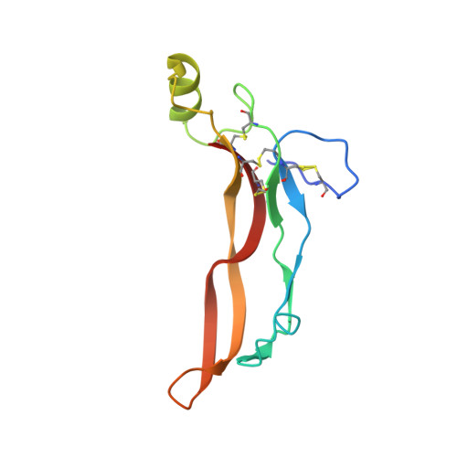
Crystal structure of human GDF11.

Padyana, A.K.Vaidialingam, B.Hayes, D.B.Gupta, P.Franti, M.Farrow, N.A.

(2016) Acta Crystallogr F Struct Biol Commun 72: 160-164

  • DOI: https://doi.org/10.1107/S2053230X16001588
  • Primary Citation Related Structures: 
    5E4G

  • PubMed Abstract: 

    Members of the TGF-β family of proteins are believed to play critical roles in cellular signaling processes such as those involved in muscle differentiation. The extent to which individual family members have been characterized and linked to biological function varies greatly. The role of myostatin, also known as growth differentiation factor 8 (GDF8), as an inhibitor of muscle differentiation is well understood through genetic linkages. In contrast, the role of growth differentiation factor 11 (GDF11) is much less well understood. In humans, the mature forms of GDF11 and myostatin are over 94% identical. In order to understand the role that the small differences in sequence may play in the differential signaling of these molecules, the crystal structure of GDF11 was determined to a resolution of 1.50 Å. A comparison of the GDF11 structure with those of other family members reveals that the canonical TGF-β domain fold is conserved. A detailed structural comparison of GDF11 and myostatin shows that several of the differences between these proteins are likely to be localized at interfaces that are critical for the interaction with downstream receptors and inhibitors.


  • Organizational Affiliation
    • Boehringer Ingelheim Pharmaceuticals, 900 Ridgebury Road, Ridgefield, CT 06877, USA.

Macromolecule Content 

  • Total Structure Weight: 12.86 kDa 
  • Atom Count: 1,069 
  • Modeled Residue Count: 108 
  • Deposited Residue Count: 109 
  • Unique protein chains: 1

Macromolecules

Find similar proteins by:|  3D Structure
Entity ID: 1
MoleculeChains  Sequence LengthOrganismDetailsImage
Growth/differentiation factor 11109Homo sapiensMutation(s): 0 
Gene Names: GDF11BMP11
UniProt & NIH Common Fund Data Resources
Find proteins for O95390 (Homo sapiens)
Explore O95390 
Go to UniProtKB:  O95390
PHAROS:  O95390
GTEx:  ENSG00000135414 
Entity Groups
Sequence Clusters30% Identity50% Identity70% Identity90% Identity95% Identity100% Identity
UniProt GroupO95390
Sequence Annotations
Expand
Reference Sequence

Experimental Data & Validation

Experimental Data

  • Method: X-RAY DIFFRACTION
  • Resolution: 1.50 Å
  • R-Value Free:  0.207 (Depositor), 0.208 (DCC) 
  • R-Value Work:  0.188 (Depositor), 0.190 (DCC) 
  • R-Value Observed: 0.189 (Depositor) 
Space Group: P 41 21 2
Unit Cell:
Length ( Å )Angle ( ˚ )
a = 32.27α = 90
b = 32.27β = 90
c = 210.602γ = 90
Software Package:
Software NamePurpose
PHENIXrefinement
PDB_EXTRACTdata extraction
d*TREKdata reduction
d*TREKdata scaling
PHASERphasing

Structure Validation

View Full Validation Report


Entry History 

Deposition Data

Revision History  (Full details and data files)

  • Version 1.0: 2016-03-09
    Type: Initial release
  • Version 1.1: 2024-11-06
    Changes: Data collection, Database references, Derived calculations, Refinement description, Structure summary