5E27 | pdb_00005e27

The structure of Resuscitation Promoting Factor B from M. tuberculosis reveals unexpected ubiquitin-like domains


Experimental Data Snapshot

  • Method: X-RAY DIFFRACTION
  • Resolution: 2.60 Å
  • R-Value Free: 
    0.291 (Depositor), 0.301 (DCC) 
  • R-Value Work: 
    0.222 (Depositor), 0.234 (DCC) 
  • R-Value Observed: 
    0.225 (Depositor) 

wwPDB Validation 3D Report Full Report

Validation slider image for 5E27

This is version 1.2 of the entry. See complete history

Literature

The structure of Resuscitation promoting factor B from M. tuberculosis reveals unexpected ubiquitin-like domains.

Ruggiero, A.Squeglia, F.Romano, M.Vitagliano, L.De Simone, A.Berisio, R.

(2015) Biochim Biophys Acta 1860: 445-451

  • DOI: https://doi.org/10.1016/j.bbagen.2015.11.001
  • Primary Citation Related Structures: 
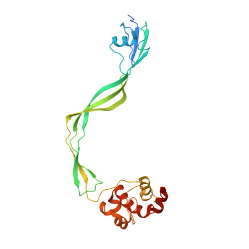
    5E27

  • PubMed Abstract: 

    RpfB is a key factor in resuscitation from dormancy of Mycobacterium tuberculosis. This protein is a cell-wall glycosidase, which cleaves cell-wall peptidoglycan. RpfB is structurally complex and is composed of three types of domains, including a catalytic, a G5 and three DUF348 domains. Structural information is currently limited to a portion of the protein including only the catalytic and G5 domains. To gain insights into the structure and function of all domains we have undertaken structural investigations on a large protein fragment containing all three types of domains that constitute RpfB (RpfB3D). The structural features of RpfB3D have been investigated combining x-ray crystallography and biophysical studies. The crystal structure of RpfB3D provides the first structural characterization of a DUF348 domain and revealed an unexpected structural relationship with ubiquitin. The crystal structure also provides specific structural features of these domains explaining their frequent association with G5 domains. Results provided novel insights into the mechanism of peptidoglycan degradation necessary to the resuscitation of M. tuberculosis. Features of the DUF348 domain add structural data to a large set of proteins embedding this domain. Based on its structural similarity to ubiquitin and frequent association to the G5 domain, we propose to name this domain as G5-linked-Ubiquitin-like domain, UBLG5.


  • Organizational Affiliation
    • Institute of Biostructures and Bioimaging, CNR, via Mezzocannone 16, Napoli, Italy.

Macromolecule Content 

  • Total Structure Weight: 51.81 kDa 
  • Atom Count: 3,387 
  • Modeled Residue Count: 446 
  • Deposited Residue Count: 496 
  • Unique protein chains: 1

Macromolecules

Find similar proteins by:|  3D Structure
Entity ID: 1
MoleculeChains  Sequence LengthOrganismDetailsImage
Resuscitation-promoting factor RpfB
A, B
248Mycobacterium tuberculosisMutation(s): 0 
Gene Names: 
EC: 3
UniProt
Find proteins for P9WG29 (Mycobacterium tuberculosis (strain ATCC 25618 / H37Rv))
Explore P9WG29 
Go to UniProtKB:  P9WG29
Entity Groups
Sequence Clusters30% Identity50% Identity70% Identity90% Identity95% Identity100% Identity
UniProt GroupP9WG29
Sequence Annotations
Expand
Reference Sequence

Experimental Data & Validation

Experimental Data

  • Method: X-RAY DIFFRACTION
  • Resolution: 2.60 Å
  • R-Value Free:  0.291 (Depositor), 0.301 (DCC) 
  • R-Value Work:  0.222 (Depositor), 0.234 (DCC) 
  • R-Value Observed: 0.225 (Depositor) 
Space Group: C 2 2 21
Unit Cell:
Length ( Å )Angle ( ˚ )
a = 102.376α = 90
b = 126.99β = 90
c = 86.286γ = 90
Software Package:
Software NamePurpose
REFMACrefinement
HKL-2000data reduction
HKL-2000data scaling
SHELXphasing

Structure Validation

View Full Validation Report


Entry History 

& Funding Information

Deposition Data


Funding OrganizationLocationGrant Number
EUGA 609535

Revision History  (Full details and data files)

  • Version 1.0: 2015-11-18
    Type: Initial release
  • Version 1.1: 2015-12-23
    Changes: Database references
  • Version 1.2: 2024-11-06
    Changes: Data collection, Database references, Structure summary