5E1H

Ricin toxin in complex with neutralizing single chain monoclonal antibodies (VHHs)


Experimental Data Snapshot

  • Method: X-RAY DIFFRACTION
  • Resolution: 2.03 Å
  • R-Value Free: 0.257 
  • R-Value Work: 0.204 
  • R-Value Observed: 0.205 

wwPDB Validation   3D Report Full Report


This is version 1.4 of the entry. See complete history


Literature

Structural analysis of nested neutralizing and non-neutralizing B cell epitopes on ricin toxin's enzymatic subunit.

Rudolph, M.J.Vance, D.J.Cassidy, M.S.Rong, Y.Shoemaker, C.B.Mantis, N.J.

(2016) Proteins 84: 1162-1172

  • DOI: https://doi.org/10.1002/prot.25062
  • Primary Citation of Related Structures:  
    4Z9K, 5E1H

  • PubMed Abstract: 

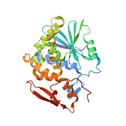
    In this report, we describe the X-ray crystal structures of two single domain camelid antibodies (VH H), F5 and F8, each in complex with ricin toxin's enzymatic subunit (RTA). F5 has potent toxin-neutralizing activity, while F8 has weak neutralizing activity. F5 buried a total of 1760 Å(2) in complex with RTA and made contact with three prominent secondary structural elements: α-helix B (Residues 98-106), β-strand h (Residues 113-117), and the C-terminus of α-helix D (Residues 154-156). F8 buried 1103 Å(2) in complex with RTA that was centered primarily on β-strand h. As such, the structural epitope of F8 is essentially nested within that of F5. All three of the F5 complementarity determining regions CDRs were involved in RTA contact, whereas F8 interactions were almost entirely mediated by CDR3, which essentially formed a seventh β-strand within RTA's centrally located β-sheet. A comparison of the two structures reported here to several previously reported (RTA-VH H) structures identifies putative contact sites on RTA, particularly α-helix B, associated with potent toxin-neutralizing activity. This information has implications for rational design of RTA-based subunit vaccines for biodefense. Proteins 2016; 84:1162-1172. © 2016 Wiley Periodicals, Inc.


  • Organizational Affiliation

    New York Structural Biology Center, New York, New York.


Macromolecules
Find similar proteins by:  (by identity cutoff)  |  3D Structure
Entity ID: 1
MoleculeChains Sequence LengthOrganismDetailsImage
Ricin258Ricinus communisMutation(s): 0 
EC: 3.2.2.22
UniProt
Find proteins for P02879 (Ricinus communis)
Explore P02879 
Go to UniProtKB:  P02879
Entity Groups  
Sequence Clusters30% Identity50% Identity70% Identity90% Identity95% Identity100% Identity
UniProt GroupP02879
Sequence Annotations
Expand
  • Reference Sequence
Find similar proteins by:  (by identity cutoff)  |  3D Structure
Entity ID: 2
MoleculeChains Sequence LengthOrganismDetailsImage
F8(JOB10) VHH antibody128Vicugna pacosMutation(s): 0 
Entity Groups  
Sequence Clusters30% Identity50% Identity70% Identity90% Identity95% Identity100% Identity
Sequence Annotations
Expand
  • Reference Sequence
Experimental Data & Validation

Experimental Data

  • Method: X-RAY DIFFRACTION
  • Resolution: 2.03 Å
  • R-Value Free: 0.257 
  • R-Value Work: 0.204 
  • R-Value Observed: 0.205 
  • Space Group: C 2 2 21
Unit Cell:
Length ( Å )Angle ( ˚ )
a = 88.472α = 90
b = 91.775β = 90
c = 95.143γ = 90
Software Package:
Software NamePurpose
PHENIXrefinement
HKL-2000data scaling
PHASERphasing
PDB_EXTRACTdata extraction
HKL-2000data reduction

Structure Validation

View Full Validation Report


Entry History & Funding Information

Deposition Data


Funding OrganizationLocationGrant Number
National Institutes of Health/National Institute Of Allergy and Infectious Diseases (NIH/NIAID)United StatesHHSN272201400021C

Revision History  (Full details and data files)

  • Version 1.0: 2016-08-03
    Type: Initial release
  • Version 1.1: 2017-04-12
    Changes: Database references, Source and taxonomy, Structure summary
  • Version 1.2: 2017-09-13
    Changes: Author supporting evidence
  • Version 1.3: 2019-12-11
    Changes: Author supporting evidence
  • Version 1.4: 2024-03-06
    Changes: Data collection, Database references