


Crystal structure of Phosphinothricin N-acetyltransferase from Brucella ovis in complex with AcetylCoA
Abendroth, J., Clifton, M.C., Lorimer, D.D., Edwards, T.E.To be published.
Experimental Data Snapshot
Starting Model: experimental
View more details
Entity ID: 1 | |||||
|---|---|---|---|---|---|
| Molecule | Chains | Sequence Length | Organism | Details | Image |
| Phosphinothricin N-acetyltransferase | 187 | Brucella ovis ATCC 25840 | Mutation(s): 0 Gene Names: pat, BOV_0087 EC: 2.3.1 | 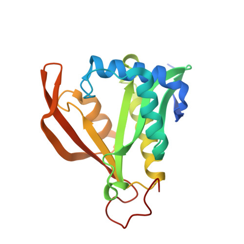 | |
UniProt | |||||
Find proteins for A0A0H3AQB6 (Brucella ovis (strain ATCC 25840 / 63/290 / NCTC 10512)) Explore A0A0H3AQB6 Go to UniProtKB: A0A0H3AQB6 | |||||
Entity Groups | |||||
| Sequence Clusters | 30% Identity50% Identity70% Identity90% Identity95% Identity100% Identity | ||||
| UniProt Group | A0A0H3AQB6 | ||||
Sequence AnnotationsExpand | |||||
Reference Sequence | |||||
| Ligands 3 Unique | |||||
|---|---|---|---|---|---|
| ID | Chains | Name / Formula / InChI Key | 2D Diagram | 3D Interactions | |
| ACO Download:Ideal Coordinates CCD File | E [auth A], G [auth B], I [auth C], J [auth D] | ACETYL COENZYME *A C23 H38 N7 O17 P3 S ZSLZBFCDCINBPY-ZSJPKINUSA-N |  | ||
| CL Download:Ideal Coordinates CCD File | F [auth A] | CHLORIDE ION Cl VEXZGXHMUGYJMC-UHFFFAOYSA-M |  | ||
| MG Download:Ideal Coordinates CCD File | H [auth B] | MAGNESIUM ION Mg JLVVSXFLKOJNIY-UHFFFAOYSA-N |  | ||
| Length ( Å ) | Angle ( ˚ ) |
|---|---|
| a = 71.1 | α = 90 |
| b = 77.38 | β = 90 |
| c = 135.84 | γ = 90 |
| Software Name | Purpose |
|---|---|
| XDS | data reduction |
| XSCALE | data scaling |
| PHASER | phasing |
| Coot | model building |
| PHENIX | refinement |
| PDB_EXTRACT | data extraction |


