5DKQ | pdb_00005dkq

Crystal Structure of Calcium-loaded S100B bound to SBi4214


Experimental Data Snapshot

  • Method: X-RAY DIFFRACTION
  • Resolution: 1.59 Å
  • R-Value Free: 
    0.230 (Depositor), 0.232 (DCC) 
  • R-Value Work: 
    0.204 (Depositor), 0.209 (DCC) 
  • R-Value Observed: 
    0.207 (Depositor) 

Starting Model: experimental
View more details

wwPDB Validation 3D Report Full Report

Validation slider image for 5DKQ

Ligand Structure Quality Assessment 


This is version 1.4 of the entry. See complete history

Literature

Small Molecule Inhibitors of Ca(2+)-S100B Reveal Two Protein Conformations.

Cavalier, M.C.Ansari, M.I.Pierce, A.D.Wilder, P.T.McKnight, L.E.Raman, E.P.Neau, D.B.Bezawada, P.Alasady, M.J.Charpentier, T.H.Varney, K.M.Toth, E.A.MacKerell, A.D.Coop, A.Weber, D.J.

(2016) J Med Chem 59: 592-608

  • DOI: https://doi.org/10.1021/acs.jmedchem.5b01369
  • Primary Citation Related Structures: 
    5DKN, 5DKQ, 5DKR

  • PubMed Abstract: 

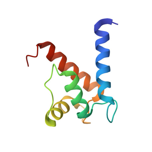
    The drug pentamidine inhibits calcium-dependent complex formation with p53 ((Ca)S100B·p53) in malignant melanoma (MM) and restores p53 tumor suppressor activity in vivo. However, off-target effects associated with this drug were problematic in MM patients. Structure-activity relationship (SAR) studies were therefore completed here with 23 pentamidine analogues, and X-ray structures of (Ca)S100B·inhibitor complexes revealed that the C-terminus of S100B adopts two different conformations, with location of Phe87 and Phe88 being the distinguishing feature and termed the "FF-gate". For symmetric pentamidine analogues ((Ca)S100B·5a, (Ca)S100B·6b) a channel between sites 1 and 2 on S100B was occluded by residue Phe88, but for an asymmetric pentamidine analogue ((Ca)S100B·17), this same channel was open. The (Ca)S100B·17 structure illustrates, for the first time, a pentamidine analog capable of binding the "open" form of the "FF-gate" and provides a means to block all three "hot spots" on (Ca)S100B, which will impact next generation (Ca)S100B·p53 inhibitor design.


  • Organizational Affiliation
    • Department of Biochemistry and Molecular Biology, Center for Biomolecular Therapeutics (CBT), University of Maryland School of Medicine , Baltimore, Maryland 21201, United States.

Macromolecule Content 

  • Total Structure Weight: 11.18 kDa 
  • Atom Count: 884 
  • Modeled Residue Count: 91 
  • Deposited Residue Count: 92 
  • Unique protein chains: 1

Macromolecules

Find similar proteins by:|  3D Structure
Entity ID: 1
MoleculeChains  Sequence LengthOrganismDetailsImage
Protein S100-B92Bos taurusMutation(s): 0 
Gene Names: S100B
UniProt
Find proteins for P02638 (Bos taurus)
Explore P02638 
Go to UniProtKB:  P02638
Entity Groups
Sequence Clusters30% Identity50% Identity70% Identity90% Identity95% Identity100% Identity
UniProt GroupP02638
Sequence Annotations
Expand
Reference Sequence

Experimental Data & Validation

Experimental Data

  • Method: X-RAY DIFFRACTION
  • Resolution: 1.59 Å
  • R-Value Free:  0.230 (Depositor), 0.232 (DCC) 
  • R-Value Work:  0.204 (Depositor), 0.209 (DCC) 
  • R-Value Observed: 0.207 (Depositor) 
Space Group: P 41 21 2
Unit Cell:
Length ( Å )Angle ( ˚ )
a = 63.485α = 90
b = 63.485β = 90
c = 48.275γ = 90
Software Package:
Software NamePurpose
Aimlessdata scaling
PHENIXrefinement
PDB_EXTRACTdata extraction
PHENIXphasing

Structure Validation

View Full Validation Report


Ligand Structure Quality Assessment 


Entry History 

& Funding Information

Deposition Data


Funding OrganizationLocationGrant Number
National Institutes of Health/National Cancer Institute (NIH/NCI)United StatesCA154274
National Institutes of Health/National Institute of General Medical Sciences (NIH/NIGMS)United StatesGM58888
National Institutes of Health/National Cancer Institute (NIH/NCI)United StatesCA107331

Revision History  (Full details and data files)

  • Version 1.0: 2016-01-20
    Type: Initial release
  • Version 1.1: 2016-02-10
    Changes: Database references
  • Version 1.2: 2017-09-27
    Changes: Author supporting evidence, Database references, Derived calculations, Refinement description
  • Version 1.3: 2019-12-04
    Changes: Author supporting evidence
  • Version 1.4: 2023-09-27
    Changes: Data collection, Database references, Refinement description