5DCW

Iridoid synthase from Catharanthus roseus - ligand free structure


Experimental Data Snapshot

  • Method: X-RAY DIFFRACTION
  • Resolution: 1.90 Å
  • R-Value Free: 0.209 
  • R-Value Work: 0.176 
  • R-Value Observed: 0.178 

wwPDB Validation   3D Report Full Report


This is version 1.3 of the entry. See complete history


Literature

Structural determinants of reductive terpene cyclization in iridoid biosynthesis.

Kries, H.Caputi, L.Stevenson, C.E.Kamileen, M.O.Sherden, N.H.Geu-Flores, F.Lawson, D.M.O'Connor, S.E.

(2016) Nat Chem Biol 12: 6-8

  • DOI: https://doi.org/10.1038/nchembio.1955
  • Primary Citation of Related Structures:  
    5DCU, 5DCW, 5DCY, 5DF1

  • PubMed Abstract: 

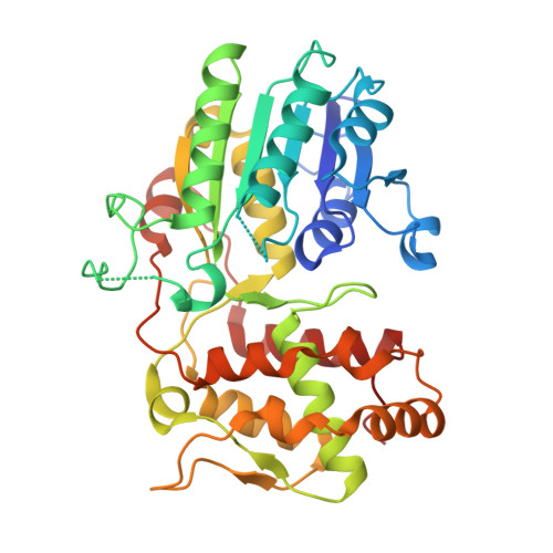
    The carbon skeleton of ecologically and pharmacologically important iridoid monoterpenes is formed in a reductive cyclization reaction unrelated to canonical terpene cyclization. Here we report the crystal structure of the recently discovered iridoid cyclase (from Catharanthus roseus) bound to a mechanism-inspired inhibitor that illuminates substrate binding and catalytic function of the enzyme. Key features that distinguish iridoid synthase from its close homolog progesterone 5β-reductase are highlighted.


  • Organizational Affiliation

    The John Innes Centre, Department of Biological Chemistry, Norwich Research Park, Norwich NR4 7UH, UK.


Macromolecules
Find similar proteins by:  (by identity cutoff)  |  3D Structure
Entity ID: 1
MoleculeChains Sequence LengthOrganismDetailsImage
Iridoid synthase371Catharanthus roseusMutation(s): 0 
EC: 1.3.1.99
UniProt
Find proteins for K7WDL7 (Catharanthus roseus)
Explore K7WDL7 
Go to UniProtKB:  K7WDL7
Entity Groups  
Sequence Clusters30% Identity50% Identity70% Identity90% Identity95% Identity100% Identity
UniProt GroupK7WDL7
Sequence Annotations
Expand
  • Reference Sequence
Experimental Data & Validation

Experimental Data

  • Method: X-RAY DIFFRACTION
  • Resolution: 1.90 Å
  • R-Value Free: 0.209 
  • R-Value Work: 0.176 
  • R-Value Observed: 0.178 
  • Space Group: H 3 2
Unit Cell:
Length ( Å )Angle ( ˚ )
a = 141.2α = 90
b = 141.2β = 90
c = 106.05γ = 120
Software Package:
Software NamePurpose
Aimlessdata scaling
REFMACrefinement
PDB_EXTRACTdata extraction
PHASERphasing
XDSdata reduction

Structure Validation

View Full Validation Report


Entry History & Funding Information

Deposition Data


Funding OrganizationLocationGrant Number
European Research CouncilUnited Kingdom311363
Swiss National Science FoundationSwitzerland155581
Biotechnology and Biological Sciences Research CouncilUnited KingdomBB/J004561/1

Revision History  (Full details and data files)

  • Version 1.0: 2015-10-28
    Type: Initial release
  • Version 1.1: 2015-11-18
    Changes: Database references
  • Version 1.2: 2015-12-30
    Changes: Database references
  • Version 1.3: 2024-01-10
    Changes: Data collection, Database references, Refinement description