5DCQ | pdb_00005dcq

Crystal structure of bacterial adhesin, FNE from Streptococcus equi spp. equi.


Experimental Data Snapshot

  • Method: X-RAY DIFFRACTION
  • Resolution: 1.83 Å
  • R-Value Free: 
    0.203 (Depositor), 0.200 (DCC) 
  • R-Value Work: 
    0.167 (Depositor), 0.163 (DCC) 
  • R-Value Observed: 
    0.169 (Depositor) 

Starting Models: experimental
View more details

wwPDB Validation 3D Report Full Report

Validation slider image for 5DCQ

This is version 1.1 of the entry. See complete history

Literature

Structural and functional analysis of the fibronectin-binding protein FNE from Streptococcus equi spp. equi.

Tiouajni, M.Durand, D.Blondeau, K.Graille, M.Urvoas, A.Valerio-Lepiniec, M.Guellouz, A.Aumont-Nicaise, M.Minard, P.van Tilbeurgh, H.

(2014) FEBS J 281: 5513-5531

  • DOI: https://doi.org/10.1111/febs.13092
  • Primary Citation Related Structures: 
    5DCQ

  • PubMed Abstract: 

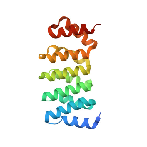
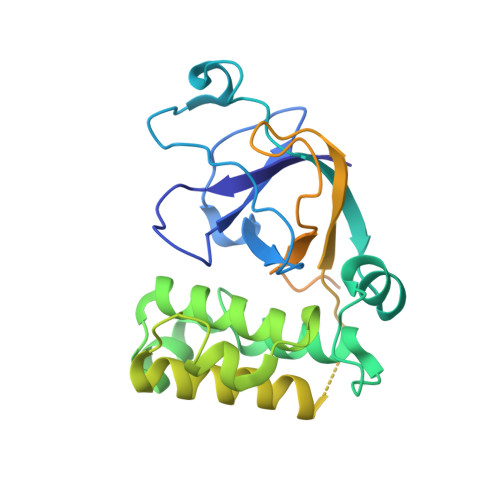
    Streptococcus equi is a horse pathogen belonging to Lancefield group C. Infection by S. equi ssp. equi causes strangles, a serious and highly contagious disease of the upper respiratory tract. S. equi ssp. equi secretes a fibronectin (Fn)-binding protein, FNE, that does not contain cell wall-anchoring motifs. FNE binds to the gelatin-binding domain (GBD) of Fn, composed of the motifs (6) FI (12) FII (789) FI . FNE lacks the canonical Fn-binding peptide repeats observed in many microbial surface components recognizing adhesive matrix molecules. We found that the interaction between FNE and the human GBD is mediated by the binding of the disordered C-terminal region (residues 208-262) of FNE to the (789) FI GBD subfragment. The crystal structure of FNE showed that it is similar to the minor pilus protein Spy0125 of Streptococcus pyogenes, found at the end of pilus polymers and responsible for adhesion. FNE and Spy0125 both have a superimposable internal thioester bond between highly conserved Cys and Gln residues. Small-angle X-ray scattering of the FNE-(789) FI complex provided a model that aligns the C-terminal peptide of FNE with the E-strands of the FI domains, adopting the β-zipper extension model observed in previous structures of microbial surface components recognizing adhesive matrix molecule adhesion peptides bound to FI domains.


  • Organizational Affiliation
    • Institut de Biochimie et de Biophysique Moléculaire et Cellulaire, UMR 8619 CNRS, Université Paris Sud, Orsay, France.

Macromolecule Content 

  • Total Structure Weight: 148.68 kDa 
  • Atom Count: 9,718 
  • Modeled Residue Count: 1,069 
  • Deposited Residue Count: 1,323 
  • Unique protein chains: 2

Macromolecules

Find similar proteins by:|  3D Structure
Entity ID: 1
MoleculeChains  Sequence LengthOrganismDetailsImage
artificial repeat proteins (alphaREP3)
A, B, C
170synthetic constructMutation(s): 0 
Find similar proteins by:|  3D Structure
Entity ID: 2
MoleculeChains  Sequence LengthOrganismDetailsImage
Fibronectin-binding protein
D, E, F
271Streptococcus equi subsp. equiMutation(s): 0 
Gene Names: fne
UniProt
Find proteins for Q93ED6 (Streptococcus equi subsp. equi)
Explore Q93ED6 
Go to UniProtKB:  Q93ED6
Entity Groups
Sequence Clusters30% Identity50% Identity70% Identity90% Identity95% Identity100% Identity
UniProt GroupQ93ED6
Sequence Annotations
Expand
Reference Sequence

Small Molecules

Ligands 1 Unique
IDChains Name / Formula / InChI Key2D Diagram3D Interactions
FMT

Query on FMT


Download:Ideal Coordinates CCD File
G [auth A]
H [auth B]
I [auth C]
J [auth C]
K [auth C]
G [auth A],
H [auth B],
I [auth C],
J [auth C],
K [auth C],
L [auth C],
M [auth D],
N [auth E],
O [auth E],
P [auth E],
Q [auth F],
R [auth F]
FORMIC ACID
C H2 O2
BDAGIHXWWSANSR-UHFFFAOYSA-N

Experimental Data & Validation

Experimental Data

  • Method: X-RAY DIFFRACTION
  • Resolution: 1.83 Å
  • R-Value Free:  0.203 (Depositor), 0.200 (DCC) 
  • R-Value Work:  0.167 (Depositor), 0.163 (DCC) 
  • R-Value Observed: 0.169 (Depositor) 
Space Group: P 61
Unit Cell:
Length ( Å )Angle ( ˚ )
a = 113.688α = 90
b = 113.688β = 90
c = 152.623γ = 120
Software Package:
Software NamePurpose
XDSdata reduction
MOLREPphasing
REFMACrefinement
PDB_EXTRACTdata extraction
XSCALEdata scaling

Structure Validation

View Full Validation Report


Entry History 

Deposition Data

Revision History  (Full details and data files)

  • Version 1.0: 2016-06-29
    Type: Initial release
  • Version 1.1: 2024-01-10
    Changes: Data collection, Database references, Refinement description