5DA4

Structure of a nanobody recognizing the fumarate transporter SLC26Dg


Experimental Data Snapshot

  • Method: X-RAY DIFFRACTION
  • Resolution: 2.40 Å
  • R-Value Free: 0.242 
  • R-Value Work: 0.233 
  • R-Value Observed: 0.233 

wwPDB Validation   3D Report Full Report


This is version 2.0 of the entry. See complete history


Literature

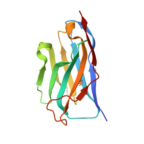
Structure of a prokaryotic fumarate transporter reveals the architecture of the SLC26 family.

Geertsma, E.R.Chang, Y.N.Shaik, F.R.Neldner, Y.Pardon, E.Steyaert, J.Dutzler, R.

(2015) Nat Struct Mol Biol 22: 803-808

  • DOI: https://doi.org/10.1038/nsmb.3091
  • Primary Citation of Related Structures:  
    5DA0, 5DA4, 5IOF

  • PubMed Abstract: 

    The SLC26 family of membrane proteins combines a variety of functions within a conserved molecular scaffold. Its members, besides coupled anion transporters and channels, include the motor protein Prestin, which confers electromotility to cochlear outer hair cells. To gain insight into the architecture of this protein family, we characterized the structure and function of SLC26Dg, a facilitator of proton-coupled fumarate symport, from the bacterium Deinococcus geothermalis. Its modular structure combines a transmembrane unit and a cytoplasmic STAS domain. The membrane-inserted domain consists of two intertwined inverted repeats of seven transmembrane segments each and resembles the fold of the unrelated transporter UraA. It shows an inward-facing, ligand-free conformation with a potential substrate-binding site at the interface between two helix termini at the center of the membrane. This structure defines the common framework for the diverse functional behavior of the SLC26 family.


  • Organizational Affiliation

    Department of Biochemistry, University of Zurich, Zurich, Switzerland.


Macromolecules
Find similar proteins by:  (by identity cutoff)  |  3D Structure
Entity ID: 1
MoleculeChains Sequence LengthOrganismDetailsImage
Nanobody recognizing the membrane protein SLC26Dg
A, B, C
125Lama glamaMutation(s): 0 
Entity Groups  
Sequence Clusters30% Identity50% Identity70% Identity90% Identity95% Identity100% Identity
Sequence Annotations
Expand
  • Reference Sequence
Experimental Data & Validation

Experimental Data

  • Method: X-RAY DIFFRACTION
  • Resolution: 2.40 Å
  • R-Value Free: 0.242 
  • R-Value Work: 0.233 
  • R-Value Observed: 0.233 
  • Space Group: P 43 2 2
Unit Cell:
Length ( Å )Angle ( ˚ )
a = 139.511α = 90
b = 139.511β = 90
c = 196.863γ = 90
Software Package:
Software NamePurpose
PHENIXrefinement
XDSdata reduction
PHENIXphasing
XSCALEdata scaling

Structure Validation

View Full Validation Report


Entry History 

Deposition Data

Revision History  (Full details and data files)

  • Version 1.0: 2015-09-09
    Type: Initial release
  • Version 1.1: 2015-09-23
    Changes: Database references
  • Version 1.2: 2015-10-14
    Changes: Database references
  • Version 2.0: 2024-01-10
    Changes: Atomic model, Data collection, Database references, Refinement description