


crystal structure of SSB from homo sapiens
Li, Y.H., Gao, Z.Q., Dong, Y.H.To be published.
Experimental Data Snapshot
wwPDB Validation 3D Report Full Report

Entity ID: 1 | |||||
|---|---|---|---|---|---|
| Molecule | Chains | Sequence Length | Organism | Details | Image |
| SOSS complex subunit B1 | 115 | Homo sapiens | Mutation(s): 1 Gene Names: NABP2, OBFC2B, SSB1, LP3587 | 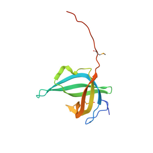 | |
UniProt & NIH Common Fund Data Resources | |||||
PHAROS: Q9BQ15 GTEx: ENSG00000139579 | |||||
Entity Groups | |||||
| Sequence Clusters | 30% Identity50% Identity70% Identity90% Identity95% Identity100% Identity | ||||
| UniProt Group | Q9BQ15 | ||||
Sequence AnnotationsExpand | |||||
Reference Sequence | |||||
| Modified Residues 1 Unique | |||||
|---|---|---|---|---|---|
| ID | Chains | Type | Formula | 2D Diagram | Parent |
| MSE Query on MSE | A, B, C, D | L-PEPTIDE LINKING | C5 H11 N O2 Se |  | MET |
| Length ( Å ) | Angle ( ˚ ) |
|---|---|
| a = 137.691 | α = 90 |
| b = 137.691 | β = 90 |
| c = 83.82 | γ = 120 |
| Software Name | Purpose |
|---|---|
| PHENIX | refinement |
| HKL-2000 | data reduction |
| HKL-2000 | data scaling |
| PHENIX | phasing |


