5D85

Staphyloferrin B precursor biosynthetic enzyme SbnA bound to aminoacrylate intermediate


Experimental Data Snapshot

  • Method: X-RAY DIFFRACTION
  • Resolution: 1.92 Å
  • R-Value Free: 0.214 
  • R-Value Work: 0.171 
  • R-Value Observed: 0.174 

wwPDB Validation   3D Report Full Report


Ligand Structure Quality Assessment 


This is version 1.4 of the entry. See complete history


Literature

Deciphering the Substrate Specificity of SbnA, the Enzyme Catalyzing the First Step in Staphyloferrin B Biosynthesis.

Kobylarz, M.J.Grigg, J.C.Liu, Y.Lee, M.S.Heinrichs, D.E.Murphy, M.E.

(2016) Biochemistry 55: 927-939

  • DOI: https://doi.org/10.1021/acs.biochem.5b01045
  • Primary Citation of Related Structures:  
    5D84, 5D85, 5D86, 5D87

  • PubMed Abstract: 

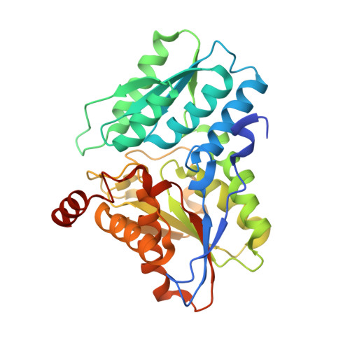
    Staphylococcus aureus assembles the siderophore, staphyloferrin B, from l-2,3-diaminopropionic acid (l-Dap), α-ketoglutarate, and citrate. Recently, SbnA and SbnB were shown to produce l-Dap and α-ketoglutarate from O-phospho-l-serine (OPS) and l-glutamate. SbnA is a pyridoxal 5'-phosphate (PLP)-dependent enzyme with homology to O-acetyl-l-serine sulfhydrylases; however, SbnA utilizes OPS instead of O-acetyl-l-serine (OAS), and l-glutamate serves as a nitrogen donor instead of a sulfide. In this work, we examined how SbnA dictates substrate specificity for OPS and l-glutamate using a combination of X-ray crystallography, enzyme kinetics, and site-directed mutagenesis. Analysis of SbnA crystals incubated with OPS revealed the structure of the PLP-α-aminoacrylate intermediate. Formation of the intermediate induced closure of the active site pocket by narrowing the channel leading to the active site and forming a second substrate binding pocket that likely binds l-glutamate. Three active site residues were identified: Arg132, Tyr152, Ser185 that were essential for OPS recognition and turnover. The Y152F/S185G SbnA double mutant was completely inactive, and its crystal structure revealed that the mutations induced a closed form of the enzyme in the absence of the α-aminoacrylate intermediate. Lastly, l-cysteine was shown to be a competitive inhibitor of SbnA by forming a nonproductive external aldimine with the PLP cofactor. These results suggest a regulatory link between siderophore and l-cysteine biosynthesis, revealing a potential mechanism to reduce iron uptake under oxidative stress.


  • Organizational Affiliation

    Department of Microbiology and Immunology, Life Sciences Institute, The University of British Columbia , Vancouver, British Columbia, Canada V6T 1Z3.


Macromolecules
Find similar proteins by:  (by identity cutoff)  |  3D Structure
Entity ID: 1
MoleculeChains Sequence LengthOrganismDetailsImage
Probable siderophore biosynthesis protein SbnA326Staphylococcus aureus subsp. aureus str. NewmanMutation(s): 0 
Gene Names: sbnANWMN_0060
UniProt
Find proteins for A6QDA0 (Staphylococcus aureus (strain Newman))
Explore A6QDA0 
Go to UniProtKB:  A6QDA0
Entity Groups  
Sequence Clusters30% Identity50% Identity70% Identity90% Identity95% Identity100% Identity
UniProt GroupA6QDA0
Sequence Annotations
Expand
  • Reference Sequence
Small Molecules
Ligands 3 Unique
IDChains Name / Formula / InChI Key2D Diagram3D Interactions
P1T
Query on P1T

Download Ideal Coordinates CCD File 
C [auth A]2-[({3-HYDROXY-2-METHYL-5-[(PHOSPHONOOXY)METHYL]PYRIDIN-4-YL}METHYL)AMINO]ACRYLIC ACID
C11 H15 N2 O7 P
BXUDKFHCAMQSRX-UHFFFAOYSA-N
FLC
Query on FLC

Download Ideal Coordinates CCD File 
B [auth A]CITRATE ANION
C6 H5 O7
KRKNYBCHXYNGOX-UHFFFAOYSA-K
GOL
Query on GOL

Download Ideal Coordinates CCD File 
D [auth A]GLYCEROL
C3 H8 O3
PEDCQBHIVMGVHV-UHFFFAOYSA-N
Experimental Data & Validation

Experimental Data

  • Method: X-RAY DIFFRACTION
  • Resolution: 1.92 Å
  • R-Value Free: 0.214 
  • R-Value Work: 0.171 
  • R-Value Observed: 0.174 
  • Space Group: P 21 21 2
Unit Cell:
Length ( Å )Angle ( ˚ )
a = 56.741α = 90
b = 117.408β = 90
c = 47.818γ = 90
Software Package:
Software NamePurpose
Aimlessdata scaling
PHENIXphasing
PHENIXrefinement
PDB_EXTRACTdata extraction
XDSdata reduction

Structure Validation

View Full Validation Report


Ligand Structure Quality Assessment 


Entry History & Funding Information

Deposition Data


Funding OrganizationLocationGrant Number
Canadian Institutes of Health Research (CIHR)CanadaMOP-102596

Revision History  (Full details and data files)

  • Version 1.0: 2016-02-03
    Type: Initial release
  • Version 1.1: 2016-02-24
    Changes: Database references
  • Version 1.2: 2017-09-06
    Changes: Author supporting evidence, Database references, Derived calculations
  • Version 1.3: 2020-01-08
    Changes: Author supporting evidence
  • Version 1.4: 2024-03-06
    Changes: Data collection, Database references