Sph3 Is a Glycoside Hydrolase Required for the Biosynthesis of Galactosaminogalactan in Aspergillus fumigatus.
Bamford, N.C., Snarr, B.D., Gravelat, F.N., Little, D.J., Lee, M.J., Zacharias, C.A., Chabot, J.C., Geller, A.M., Baptista, S.D., Baker, P., Robinson, H., Howell, P.L., Sheppard, D.C.(2015) J Biological Chem 290: 27438-27450
- PubMed: 26342082 Search on PubMedSearch on PubMed Central
- DOI: https://doi.org/10.1074/jbc.M115.679050
- Primary Citation Related Structures:
5C5G, 5D6T - PubMed Abstract:
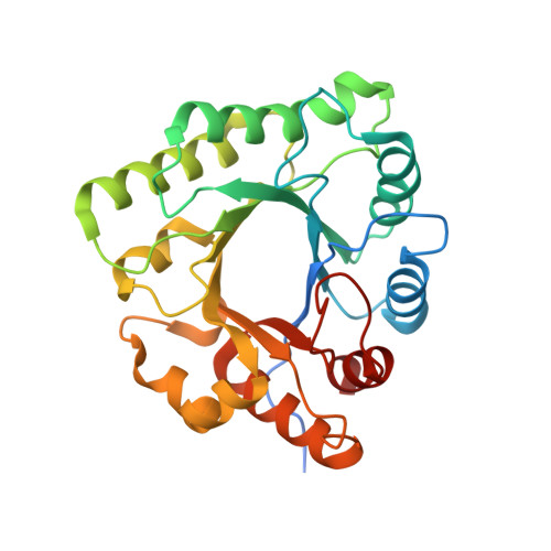
Aspergillus fumigatus is the most virulent species within the Aspergillus genus and causes invasive infections with high mortality rates. The exopolysaccharide galactosaminogalactan (GAG) contributes to the virulence of A. fumigatus. A co-regulated five-gene cluster has been identified and proposed to encode the proteins required for GAG biosynthesis. One of these genes, sph3, is predicted to encode a protein belonging to the spherulin 4 family, a protein family with no known function. Construction of an sph3-deficient mutant demonstrated that the gene is necessary for GAG production. To determine the role of Sph3 in GAG biosynthesis, we determined the structure of Aspergillus clavatus Sph3 to 1.25 Å. The structure revealed a (β/α)8 fold, with similarities to glycoside hydrolase families 18, 27, and 84. Recombinant Sph3 displayed hydrolytic activity against both purified and cell wall-associated GAG. Structural and sequence alignments identified three conserved acidic residues, Asp-166, Glu-167, and Glu-222, that are located within the putative active site groove. In vitro and in vivo mutagenesis analysis demonstrated that all three residues are important for activity. Variants of Asp-166 yielded the greatest decrease in activity suggesting a role in catalysis. This work shows that Sph3 is a glycoside hydrolase essential for GAG production and defines a new glycoside hydrolase family, GH135.
- From the Program in Molecular Structure and Function, Research Institute, The Hospital for Sick Children, Toronto, Ontario M5G 0A4, Canada, the Department of Biochemistry, University of Toronto, Toronto, Ontario M5S 1A8, Canada.
Organizational Affiliation: