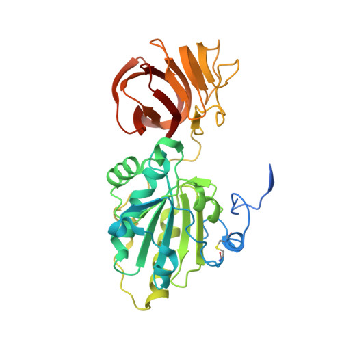
Structure of the ectodomain of the electron transporter Rv2874 from Mycobacterium tuberculosis reveals a thioredoxin-like domain combined with a carbohydrate-binding module.
Goldstone, D.C., Metcalf, P., Baker, E.N.(2016) Acta Crystallogr D Struct Biol 72: 40-48
- PubMed: 26894533 Search on PubMed
- DOI: https://doi.org/10.1107/S2059798315021488
- Primary Citation Related Structures:
5CYY - PubMed Abstract:
The members of the CcdA family are integral membrane proteins that use a disulfide cascade to transport electrons from the thioredoxin-thioredoxin reductase system in the interior of the cell into the extracytoplasmic space. The core transmembrane portion of this family is often elaborated with additional hydrophilic domains that act as adapters to deliver reducing potential to targets outside the cellular membrane. To investigate the function of family members in Mycobacterium tuberculosis, the structure of the C-terminal ectodomain from Rv2874, one of three CcdA-family members present in the genome, was determined. The crystal structure, which was refined at 1.9 Å resolution with R = 0.195 and Rfree = 0.219, reveals the predicted thioredoxin-like domain with its conserved Cys-X-X-Cys active-site motif. Unexpectedly, this domain is combined with a second domain with a carbohydrate-binding module (CBM) fold, this being the first reported example of a CBM in association with a thioredoxin-like domain fold. A cavity in the CBM adjacent to the thioredoxin active site suggests a likely carbohydrate-binding site, representing a broadening of the substrate range for CcdA-family members and an expansion of the thioredoxin-domain functionality to carbohydrate modification.
- School of Biological Sciences, University of Auckland, Private Bag 92-019, Auckland, New Zealand.
Organizational Affiliation: