


Structure of sinapyl alcohol bound monolignol 4-O-methyltransferase at 1.60 Angstroms resolution
Cai, Y., Liu, C.-J.To be published.
Experimental Data Snapshot
Entity ID: 1 | |||||
|---|---|---|---|---|---|
| Molecule | Chains | Sequence Length | Organism | Details | Image |
| (Iso)eugenol O-methyltransferase | 368 | Clarkia breweri | Mutation(s): 5 Gene Names: IEMT1 EC: 2.1.1.146 | 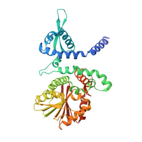 | |
UniProt | |||||
Entity Groups | |||||
| Sequence Clusters | 30% Identity50% Identity70% Identity90% Identity95% Identity100% Identity | ||||
| UniProt Group | O04385 | ||||
Sequence AnnotationsExpand | |||||
Reference Sequence | |||||
| Ligands 3 Unique | |||||
|---|---|---|---|---|---|
| ID | Chains | Name / Formula / InChI Key | 2D Diagram | 3D Interactions | |
| SAH Download:Ideal Coordinates CCD File | F [auth A], K [auth B], N [auth C], S [auth D] | S-ADENOSYL-L-HOMOCYSTEINE C14 H20 N6 O5 S ZJUKTBDSGOFHSH-WFMPWKQPSA-N |  | ||
| 55B Download:Ideal Coordinates CCD File | E [auth A], J [auth B], M [auth C], R [auth D] | 4-[(1E)-3-hydroxyprop-1-en-1-yl]-2,6-dimethoxyphenol C11 H14 O4 LZFOPEXOUVTGJS-ONEGZZNKSA-N |  | ||
| NO3 Download:Ideal Coordinates CCD File | G [auth A] H [auth A] I [auth A] L [auth B] O [auth C] | NITRATE ION N O3 NHNBFGGVMKEFGY-UHFFFAOYSA-N |  | ||
| Length ( Å ) | Angle ( ˚ ) |
|---|---|
| a = 66.54 | α = 90 |
| b = 151.63 | β = 92.55 |
| c = 68.39 | γ = 90 |
| Software Name | Purpose |
|---|---|
| REFMAC | refinement |
| MOSFLM | data reduction |
| SCALA | data scaling |
| MOLREP | phasing |
| Funding Organization | Location | Grant Number |
|---|---|---|
| Department of Energy (DOE, United States) | United States | DEAC0298CH10886 |


