5CFY | pdb_00005cfy

CRYSTAL STRUCTURE OF GLTPH R397A IN COMPLEX WITH NA+ AND L-ASP


Experimental Data Snapshot

  • Method: X-RAY DIFFRACTION
  • Resolution: 3.50 Å
  • R-Value Free: 
    0.294 (Depositor), 0.289 (DCC) 
  • R-Value Work: 
    0.249 (Depositor), 0.249 (DCC) 
  • R-Value Observed: 
    0.251 (Depositor) 

Starting Model: experimental
View more details

wwPDB Validation 3D Report Full Report

Validation slider image for 5CFY

This is version 1.1 of the entry. See complete history

Literature

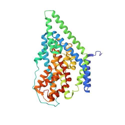
Coupled ion binding and structural transitions along the transport cycle of glutamate transporters.

Verdon, G.Oh, S.Serio, R.N.Boudker, O.

(2014) Elife 3: e02283-e02283

  • DOI: https://doi.org/10.7554/eLife.02283
  • Primary Citation Related Structures: 
    4OYE, 4OYF, 4P19, 4P1A, 4P3J, 4P6H, 5CFY

  • PubMed Abstract: 

    Membrane transporters that clear the neurotransmitter glutamate from synapses are driven by symport of sodium ions and counter-transport of a potassium ion. Previous crystal structures of a homologous archaeal sodium and aspartate symporter showed that a dedicated transport domain carries the substrate and ions across the membrane. Here, we report new crystal structures of this homologue in ligand-free and ions-only bound outward- and inward-facing conformations. We show that after ligand release, the apo transport domain adopts a compact and occluded conformation that can traverse the membrane, completing the transport cycle. Sodium binding primes the transport domain to accept its substrate and triggers extracellular gate opening, which prevents inward domain translocation until substrate binding takes place. Furthermore, we describe a new cation-binding site ideally suited to bind a counter-transported ion. We suggest that potassium binding at this site stabilizes the translocation-competent conformation of the unloaded transport domain in mammalian homologues.DOI: http://dx.doi.org/10.7554/eLife.02283.001.


  • Organizational Affiliation
    • Department of Physiology and Biophysics, Weill Cornell Medical College, New York, United States g.verdon@imperial.ac.uk.

Macromolecule Content 

  • Total Structure Weight: 270 kDa 
  • Atom Count: 18,240 
  • Modeled Residue Count: 2,466 
  • Deposited Residue Count: 2,550 
  • Unique protein chains: 1

Macromolecules

Find similar proteins by:|  3D Structure
Entity ID: 1
MoleculeChains  Sequence LengthOrganismDetailsImage
425aa long hypothetical proton glutamate symport protein
A, B, C, D, E
A, B, C, D, E, F
425Pyrococcus horikoshii OT3Mutation(s): 7 
Gene Names: PH1295
Membrane Entity: Yes 
UniProt
Find proteins for O59010 (Pyrococcus horikoshii (strain ATCC 700860 / DSM 12428 / JCM 9974 / NBRC 100139 / OT-3))
Explore O59010 
Go to UniProtKB:  O59010
Entity Groups
Sequence Clusters30% Identity50% Identity70% Identity90% Identity95% Identity100% Identity
UniProt GroupO59010
Sequence Annotations
Expand
Reference Sequence

Small Molecules

Ligands 2 Unique
IDChains Name / Formula / InChI Key2D Diagram3D Interactions
ASP

Query on ASP


Download:Ideal Coordinates CCD File
G [auth A]
J [auth B]
M [auth C]
P [auth D]
S [auth E]
G [auth A],
J [auth B],
M [auth C],
P [auth D],
S [auth E],
V [auth F]
ASPARTIC ACID
C4 H7 N O4
CKLJMWTZIZZHCS-REOHCLBHSA-N
NA

Query on NA


Download:Ideal Coordinates CCD File
H [auth A]
I [auth A]
K [auth B]
L [auth B]
N [auth C]
H [auth A],
I [auth A],
K [auth B],
L [auth B],
N [auth C],
O [auth C],
Q [auth D],
R [auth D],
T [auth E],
U [auth E],
W [auth F],
X [auth F]
SODIUM ION
Na
FKNQFGJONOIPTF-UHFFFAOYSA-N

Experimental Data & Validation

Experimental Data

  • Method: X-RAY DIFFRACTION
  • Resolution: 3.50 Å
  • R-Value Free:  0.294 (Depositor), 0.289 (DCC) 
  • R-Value Work:  0.249 (Depositor), 0.249 (DCC) 
  • R-Value Observed: 0.251 (Depositor) 
Space Group: P 31
Unit Cell:
Length ( Å )Angle ( ˚ )
a = 116.958α = 90
b = 116.958β = 90
c = 313.517γ = 120
Software Package:
Software NamePurpose
REFMACrefinement
HKL-2000data reduction
HKL-2000data scaling
PHASERphasing
Cootmodel building

Structure Validation

View Full Validation Report


Entry History 

Deposition Data

Revision History  (Full details and data files)

  • Version 1.0: 2016-04-20
    Type: Initial release
  • Version 1.1: 2023-09-27
    Changes: Data collection, Database references, Derived calculations, Refinement description