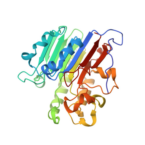
Structural comparison of AP endonucleases from the exonuclease III family reveals new amino acid residues in human AP endonuclease 1 that are involved in incision of damaged DNA.
Redrejo-Rodriguez, M., Vigouroux, A., Mursalimov, A., Grin, I., Alili, D., Koshenov, Z., Akishev, Z., Maksimenko, A., Bissenbaev, A.K., Matkarimov, B.T., Saparbaev, M., Ishchenko, A.A., Morera, S.(2016) Biochimie 128-129: 20-33
- PubMed: 27343627 Search on PubMed
- DOI: https://doi.org/10.1016/j.biochi.2016.06.011
- Primary Citation Related Structures:
5CFE, 5CFG - PubMed Abstract:
Oxidatively damaged DNA bases are substrates for two overlapping repair pathways: DNA glycosylase-initiated base excision repair (BER) and apurinic/apyrimidinic (AP) endonuclease-initiated nucleotide incision repair (NIR). In the BER pathway, an AP endonuclease cleaves DNA at AP sites and 3'-blocking moieties generated by DNA glycosylases, whereas in the NIR pathway, the same AP endonuclease incises DNA 5' to an oxidized base. The majority of characterized AP endonucleases possess classic BER activities, and approximately a half of them can also have a NIR activity. At present, the molecular mechanism underlying DNA substrate specificity of AP endonucleases remains unclear mainly due to the absence of a published structure of the enzyme in complex with a damaged base. To identify critical residues involved in the NIR function, we performed biochemical and structural characterization of Bacillus subtilis AP endonuclease ExoA and compared its crystal structure with the structures of other AP endonucleases: Escherichia coli exonuclease III (Xth), human APE1, and archaeal Mth212. We found conserved amino acid residues in the NIR-specific enzymes APE1, Mth212, and ExoA. Four of these positions were studied by means of point mutations in APE1: we applied substitution with the corresponding residue found in NIR-deficient E. coli Xth (Y128H, N174Q, G231S, and T268D). The APE1-T268D mutant showed a drastically decreased NIR activity and an inverted Mg(2+) dependence of the AP site cleavage activity, which is in line with the presence of an aspartic residue at the equivalent position among other known NIR-deficient AP endonucleases. Taken together, these data show that NIR is an evolutionarily conserved function in the Xth family of AP endonucleases.
- Laboratoire «Stabilité Génétique et Oncogenèse» CNRS, UMR 8200, Univ. Paris-Sud, Université Paris-Saclay, Gustave Roussy Cancer Campus, Equipe Labellisée Ligue Contre le Cancer, F-94805 Villejuif Cedex, France.
Organizational Affiliation: