High-resolution neutron and X-ray diffraction room-temperature studies of an H-FABP-oleic acid complex: study of the internal water cluster and ligand binding by a transferred multipolar electron-density distribution.
Howard, E.I., Guillot, B., Blakeley, M.P., Haertlein, M., Moulin, M., Mitschler, A., Cousido-Siah, A., Fadel, F., Valsecchi, W.M., Tomizaki, T., Petrova, T., Claudot, J., Podjarny, A.(2016) IUCrJ 3: 115-126
- PubMed: 27006775 Search on PubMedSearch on PubMed Central
- DOI: https://doi.org/10.1107/S2052252515024161
- Primary Citation Related Structures:
5CE4 - PubMed Abstract:
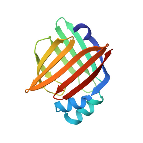
Crystal diffraction data of heart fatty acid binding protein (H-FABP) in complex with oleic acid were measured at room temperature with high-resolution X-ray and neutron protein crystallography (0.98 and 1.90 Å resolution, respectively). These data provided very detailed information about the cluster of water molecules and the bound oleic acid in the H-FABP large internal cavity. The jointly refined X-ray/neutron structure of H-FABP was complemented by a transferred multipolar electron-density distribution using the parameters of the ELMAMII library. The resulting electron density allowed a precise determination of the electrostatic potential in the fatty acid (FA) binding pocket. Bader's quantum theory of atoms in molecules was then used to study interactions involving the internal water molecules, the FA and the protein. This approach showed H⋯H contacts of the FA with highly conserved hydrophobic residues known to play a role in the stabilization of long-chain FAs in the binding cavity. The determination of water hydrogen (deuterium) positions allowed the analysis of the orientation and electrostatic properties of the water molecules in the very ordered cluster. As a result, a significant alignment of the permanent dipoles of the water molecules with the protein electrostatic field was observed. This can be related to the dielectric properties of hydration layers around proteins, where the shielding of electrostatic interactions depends directly on the rotational degrees of freedom of the water molecules in the interface.
- Department of Integrative Biology, Institut de Génétique et de Biologie Moléculaire et Cellulaire, Centre de Biologie Intégrative, CNRS, INSERM, UdS, 1 rue Laurent Fries, 67404 Illkirch CEDEX, France; Instituto de Fisica de Liquidos y Sistemas Biologicos, CONICET, UNLP, Calle 59 No. 789, La Plata, Argentina.
Organizational Affiliation: