5CDG | pdb_00005cdg

I220F horse liver alcohol dehydrogenase complexed with NAD and pentafluorobenzyl alcohol


Experimental Data Snapshot

  • Method: X-RAY DIFFRACTION
  • Resolution: 1.40 Å
  • R-Value Free: 
    0.187 (Depositor), 0.190 (DCC) 
  • R-Value Work: 
    0.142 (Depositor), 0.148 (DCC) 
  • R-Value Observed: 
    0.142 (Depositor) 

Starting Model: experimental
View more details

wwPDB Validation 3D Report Full Report

Validation slider image for 5CDG

Ligand Structure Quality Assessment 


This is version 1.4 of the entry. See complete history

Literature

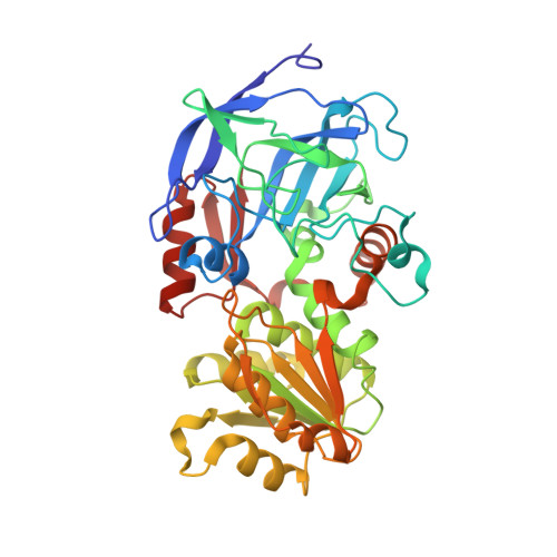
Contribution of Buried Distal Amino Acid Residues in Horse Liver Alcohol Dehydrogenase to Structure and Catalysis.

Shanmuganatham, K.K.Wallace, R.S.Lee, A.T.Plapp, B.V.

(2017) Protein Sci 

  • DOI: https://doi.org/10.1002/pro.3370
  • Primary Citation Related Structures: 
    5CDG, 5CDS, 5CDT, 5CDU, 5KJ6, 5KJC, 5KJE, 5KJF

  • PubMed Abstract: 

    The dynamics of enzyme catalysis range from the slow time scale (∼ms) for substrate binding and conformational changes to the fast time (∼ps) scale for reorganization of substrates in the chemical step. The contribution of global dynamics to catalysis by alcohol dehydrogenase was tested by substituting five different, conserved amino acid residues that are distal from the active site and located in the hinge region for the conformational change or in hydrophobic clusters. X-ray crystallography shows that the structures for the G173A, V197I, I220 (V, L, or F), V222I, and F322L enzymes complexed with NAD + and an analogue of benzyl alcohol are almost identical, except for small perturbations at the sites of substitution. The enzymes have very similar kinetic constants for the oxidation of benzyl alcohol and reduction of benzaldehyde as compared to the wild-type enzyme, and the rates of conformational changes are not altered. Less conservative substitutions of these amino acid residues, such as G173(V, E, K, or R), V197(G, S, or T), I220(G, S, T, or N), and V222(G, S, or T) produced unstable or poorly expressed proteins, indicating that the residues are critical for global stability. The enzyme scaffold accommodates conservative substitutions of distal residues, and there is no evidence that fast, global dynamics significantly affect the rate constants for hydride transfers. In contrast, other studies show that proximal residues significantly participate in catalysis.


  • Organizational Affiliation
    • Department of Biochemistry, The University of Iowa, Iowa City, IA, 52242-1109.

Macromolecule Content 

  • Total Structure Weight: 82 kDa 
  • Atom Count: 6,735 
  • Modeled Residue Count: 748 
  • Deposited Residue Count: 748 
  • Unique protein chains: 1

Macromolecules

Find similar proteins by:|  3D Structure
Entity ID: 1
MoleculeChains  Sequence LengthOrganismDetailsImage
Alcohol dehydrogenase E chain
A, B
374Equus caballusMutation(s): 1 
EC: 1.1.1.1
UniProt
Find proteins for P00327 (Equus caballus)
Explore P00327 
Go to UniProtKB:  P00327
Entity Groups
Sequence Clusters30% Identity50% Identity70% Identity90% Identity95% Identity100% Identity
UniProt GroupP00327
Sequence Annotations
Expand
Reference Sequence

Small Molecules

Ligands 4 Unique
IDChains Name / Formula / InChI Key2D Diagram3D Interactions
NAJ

Query on NAJ


Download:Ideal Coordinates CCD File
E [auth A],
K [auth B]
NICOTINAMIDE-ADENINE-DINUCLEOTIDE (ACIDIC FORM)
C21 H27 N7 O14 P2
BAWFJGJZGIEFAR-NNYOXOHSSA-N
PFB

Query on PFB


Download:Ideal Coordinates CCD File
F [auth A],
L [auth B]
2,3,4,5,6-PENTAFLUOROBENZYL ALCOHOL
C7 H3 F5 O
PGJYYCIOYBZTPU-UHFFFAOYSA-N
MRD

Query on MRD


Download:Ideal Coordinates CCD File
G [auth A],
H [auth A]
(4R)-2-METHYLPENTANE-2,4-DIOL
C6 H14 O2
SVTBMSDMJJWYQN-RXMQYKEDSA-N
ZN

Query on ZN


Download:Ideal Coordinates CCD File
C [auth A],
D [auth A],
I [auth B],
J [auth B]
ZINC ION
Zn
PTFCDOFLOPIGGS-UHFFFAOYSA-N

Experimental Data & Validation

Experimental Data

  • Method: X-RAY DIFFRACTION
  • Resolution: 1.40 Å
  • R-Value Free:  0.187 (Depositor), 0.190 (DCC) 
  • R-Value Work:  0.142 (Depositor), 0.148 (DCC) 
  • R-Value Observed: 0.142 (Depositor) 
Space Group: P 1
Unit Cell:
Length ( Å )Angle ( ˚ )
a = 44.26α = 91.75
b = 51.38β = 103.1
c = 92.39γ = 109.36
Software Package:
Software NamePurpose
REFMACrefinement
d*TREKdata reduction
d*TREKdata scaling
Omodel building
REFMACphasing

Structure Validation

View Full Validation Report


Ligand Structure Quality Assessment 


Entry History 

& Funding Information

Deposition Data


Funding OrganizationLocationGrant Number
National Institutes of Health/National Institute of General Medical Sciences (NIH/NIGMS)United StatesGM 078446

Revision History  (Full details and data files)

  • Version 1.0: 2015-07-15
    Type: Initial release
  • Version 1.1: 2017-09-27
    Changes: Author supporting evidence, Derived calculations, Source and taxonomy
  • Version 1.2: 2018-01-03
    Changes: Database references
  • Version 1.3: 2019-12-25
    Changes: Author supporting evidence
  • Version 1.4: 2023-09-27
    Changes: Data collection, Database references, Derived calculations, Refinement description