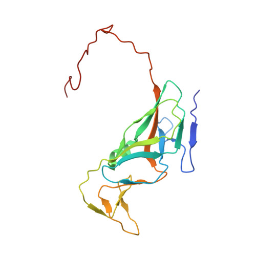
5CCT

Staphylococcus bacteriophage 80alpha dUTPase G164S mutant with dUpNHpp.


Experimental Data Snapshot

  • Method: X-RAY DIFFRACTION
  • Resolution: 2.40 Å
  • R-Value Free: 0.249 
  • R-Value Work: 0.216 
  • R-Value Observed: 0.217 

wwPDB Validation   3D Report Full Report


Ligand Structure Quality Assessment 


This is version 1.3 of the entry. See complete history


Literature

Another look at the mechanism involving trimeric dUTPases in Staphylococcus aureus pathogenicity island induction involves novel players in the party.

Maiques, E.Quiles-Puchalt, N.Donderis, J.Ciges-Tomas, J.R.Alite, C.Bowring, J.Z.Humphrey, S.Penades, J.R.Marina, A.

(2016) Nucleic Acids Res 44: 5457-5469

  • DOI: https://doi.org/10.1093/nar/gkw317
  • Primary Citation of Related Structures:  
    5CCO, 5CCT

  • PubMed Abstract: 

    We have recently proposed that the trimeric staphylococcal phage encoded dUTPases (Duts) are signaling molecules that act analogously to eukaryotic G-proteins, using dUTP as a second messenger. To perform this regulatory role, the Duts require their characteristic extra motif VI, present in all the staphylococcal phage coded trimeric Duts, as well as the strongly conserved Dut motif V. Recently, however, an alternative model involving Duts in the transfer of the staphylococcal islands (SaPIs) has been suggested, questioning the implication of motifs V and VI. Here, using state-of the-art techniques, we have revisited the proposed models. Our results confirm that the mechanism by which the Duts derepress the SaPI cycle depends on dUTP and involves both motifs V and VI, as we have previously proposed. Surprisingly, the conserved Dut motif IV is also implicated in SaPI derepression. However, and in agreement with the proposed alternative model, the dUTP inhibits rather than inducing the process, as we had initially proposed. In summary, our results clarify, validate and establish the mechanism by which the Duts perform regulatory functions.


  • Organizational Affiliation

    Instituto de Biomedicina de Valencia (IBV-CSIC) and CIBER de Enfermedades Raras (CIBERER), 46010 Valencia, Spain.


Macromolecules
Find similar proteins by:  (by identity cutoff)  |  3D Structure
Entity ID: 1
MoleculeChains Sequence LengthOrganismDetailsImage
DUTPase170Dubowvirus dv80alphaMutation(s): 1 
UniProt
Find proteins for A4ZF98 (Dubowvirus dv80alpha)
Explore A4ZF98 
Go to UniProtKB:  A4ZF98
Entity Groups  
Sequence Clusters30% Identity50% Identity70% Identity90% Identity95% Identity100% Identity
UniProt GroupA4ZF98
Sequence Annotations
Expand
  • Reference Sequence
Experimental Data & Validation

Experimental Data

  • Method: X-RAY DIFFRACTION
  • Resolution: 2.40 Å
  • R-Value Free: 0.249 
  • R-Value Work: 0.216 
  • R-Value Observed: 0.217 
  • Space Group: P 21 3
Unit Cell:
Length ( Å )Angle ( ˚ )
a = 87.464α = 90
b = 87.464β = 90
c = 87.464γ = 90
Software Package:
Software NamePurpose
PHENIXrefinement
XDSdata reduction
MOLREPphasing
SCALAdata scaling

Structure Validation

View Full Validation Report


Ligand Structure Quality Assessment 


Entry History & Funding Information

Deposition Data


Funding OrganizationLocationGrant Number
Ministerio de Ciencia e InnovacionSpainBIO2013-42619-P
Medical Research Council (United Kingdom)United KingdomMR/M003876/1
European Research CouncilERC-ADG-2014 Proposal n 670932

Revision History  (Full details and data files)

  • Version 1.0: 2016-05-11
    Type: Initial release
  • Version 1.1: 2016-06-29
    Changes: Database references
  • Version 1.2: 2017-09-13
    Changes: Author supporting evidence
  • Version 1.3: 2024-01-10
    Changes: Data collection, Database references, Derived calculations, Refinement description