5CA6

Crystallographic structure of apo porcine rotavirus TFR-41 VP8*


Experimental Data Snapshot

  • Method: X-RAY DIFFRACTION
  • Resolution: 1.90 Å
  • R-Value Free: 0.203 
  • R-Value Work: 0.164 
  • R-Value Observed: 0.166 

wwPDB Validation   3D Report Full Report


Ligand Structure Quality Assessment 


This is version 1.3 of the entry. See complete history


Literature

Substantial Receptor-induced Structural Rearrangement of Rotavirus VP8*: Potential Implications for Cross-Species Infection.

Yu, X.Mishra, R.Holloway, G.von Itzstein, M.Coulson, B.S.Blanchard, H.

(2015) Chembiochem 16: 2176-2181

  • DOI: https://doi.org/10.1002/cbic.201500360
  • Primary Citation of Related Structures:  
    5CA6, 5CAZ, 5CB7

  • PubMed Abstract: 

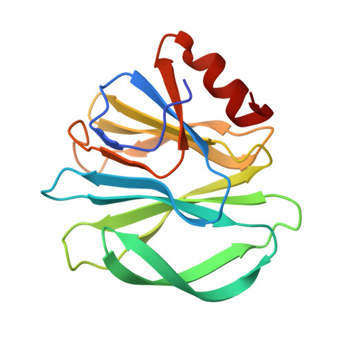
    Rotavirus-cell binding is the essential first step in rotavirus infection. This binding is a major determinant of rotavirus tropism, as host cell invasion is necessary to initiate infection. Initial rotavirus-cell interactions are mediated by carbohydrate-recognizing domain VP8* of the rotavirus capsid spike protein VP4. Here, we report the first observation of significant structural rearrangement of VP8* from human and animal rotavirus strains upon glycan receptor binding. The structural adaptability of rotavirus VP8* delivers important insights into how human and animal rotaviruses utilize the wider range of cellular glycans identified as VP8* binding partners. Furthermore, our studies on rotaviruses with atypical genetic makeup provide information expected to be critical for understanding the mechanisms of animal rotavirus gene emergence in humans and support implementation of epidemiologic surveillance of animal reservoirs as well as future vaccination schemes.


  • Organizational Affiliation

    Institute for Glycomics, Griffith University Gold Coast Campus, Southport, QLD, 4222, Australia. h.blanchard@griffith.edu.au.


Macromolecules
Find similar proteins by:  (by identity cutoff)  |  3D Structure
Entity ID: 1
MoleculeChains Sequence LengthOrganismDetailsImage
porcine rotavirus TFR-41 VP8*
A, B
161Porcine rotavirus serotype 5/strain TFR-41Mutation(s): 0 
UniProt
Find proteins for A0A1A9TAH8 (Rotavirus A (isolate RVA/Pig/Australia/TFR-41/1987/G5P9[7]))
Explore A0A1A9TAH8 
Go to UniProtKB:  A0A1A9TAH8
Entity Groups  
Sequence Clusters30% Identity50% Identity70% Identity90% Identity95% Identity100% Identity
UniProt GroupA0A1A9TAH8
Sequence Annotations
Expand
  • Reference Sequence
Small Molecules
Ligands 4 Unique
IDChains Name / Formula / InChI Key2D Diagram3D Interactions
VCA
Query on VCA

Download Ideal Coordinates CCD File 
N [auth B]VACCENIC ACID
C18 H34 O2
UWHZIFQPPBDJPM-FPLPWBNLSA-N
PLM
Query on PLM

Download Ideal Coordinates CCD File 
I [auth A]PALMITIC ACID
C16 H32 O2
IPCSVZSSVZVIGE-UHFFFAOYSA-N
GOL
Query on GOL

Download Ideal Coordinates CCD File 
C [auth A]
D [auth A]
E [auth A]
J [auth B]
K [auth B]
C [auth A],
D [auth A],
E [auth A],
J [auth B],
K [auth B],
L [auth B]
GLYCEROL
C3 H8 O3
PEDCQBHIVMGVHV-UHFFFAOYSA-N
FMT
Query on FMT

Download Ideal Coordinates CCD File 
F [auth A],
G [auth A],
H [auth A],
M [auth B]
FORMIC ACID
C H2 O2
BDAGIHXWWSANSR-UHFFFAOYSA-N
Experimental Data & Validation

Experimental Data

  • Method: X-RAY DIFFRACTION
  • Resolution: 1.90 Å
  • R-Value Free: 0.203 
  • R-Value Work: 0.164 
  • R-Value Observed: 0.166 
  • Space Group: C 1 2 1
Unit Cell:
Length ( Å )Angle ( ˚ )
a = 134.816α = 90
b = 30.444β = 124.05
c = 86.217γ = 90
Software Package:
Software NamePurpose
REFMACrefinement
XDSdata reduction
SCALAdata scaling
PHASERphasing

Structure Validation

View Full Validation Report


Ligand Structure Quality Assessment 


Entry History & Funding Information

Deposition Data


Funding OrganizationLocationGrant Number
National Health and Medical Research Council (NHMRC, Australia)AustraliaAPP1085596

Revision History  (Full details and data files)

  • Version 1.0: 2016-06-08
    Type: Initial release
  • Version 1.1: 2017-09-20
    Changes: Author supporting evidence, Data collection, Derived calculations
  • Version 1.2: 2020-01-08
    Changes: Author supporting evidence
  • Version 1.3: 2023-09-27
    Changes: Data collection, Database references, Refinement description