5BXV

eIF4E complex


Experimental Data Snapshot

  • Method: X-RAY DIFFRACTION
  • Resolution: 2.10 Å
  • R-Value Free: 0.195 
  • R-Value Work: 0.162 
  • R-Value Observed: 0.163 

wwPDB Validation   3D Report Full Report


Ligand Structure Quality Assessment 


This is version 1.6 of the entry. See complete history


Literature

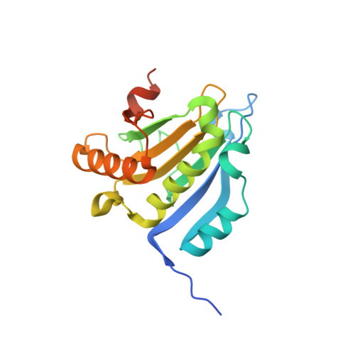
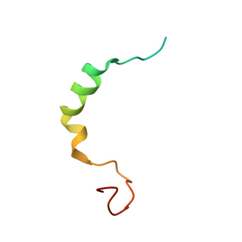
Molecular mechanism of the dual activity of 4EGI-1: Dissociating eIF4G from eIF4E but stabilizing the binding of unphosphorylated 4E-BP1.

Sekiyama, N.Arthanari, H.Papadopoulos, E.Rodriguez-Mias, R.A.Wagner, G.Leger-Abraham, M.

(2015) Proc Natl Acad Sci U S A 112: E4036-E4045

  • DOI: https://doi.org/10.1073/pnas.1512118112
  • Primary Citation of Related Structures:  
    5BXV

  • PubMed Abstract: 

    The eIF4E-binding protein (4E-BP) is a phosphorylation-dependent regulator of protein synthesis. The nonphosphorylated or minimally phosphorylated form binds translation initiation factor 4E (eIF4E), preventing binding of eIF4G and the recruitment of the small ribosomal subunit. Signaling events stimulate serial phosphorylation of 4E-BP, primarily by mammalian target of rapamycin complex 1 (mTORC1) at residues T37/T46, followed by T70 and S65. Hyperphosphorylated 4E-BP dissociates from eIF4E, allowing eIF4E to interact with eIF4G and translation initiation to resume. Because overexpression of eIF4E is linked to cellular transformation, 4E-BP is a tumor suppressor, and up-regulation of its activity is a goal of interest for cancer therapy. A recently discovered small molecule, eIF4E/eIF4G interaction inhibitor 1 (4EGI-1), disrupts the eIF4E/eIF4G interaction and promotes binding of 4E-BP1 to eIF4E. Structures of 14- to 16-residue 4E-BP fragments bound to eIF4E contain the eIF4E consensus binding motif, (54)YXXXXLΦ(60) (motif 1) but lack known phosphorylation sites. We report here a 2.1-Å crystal structure of mouse eIF4E in complex with m(7)GTP and with a fragment of human 4E-BP1, extended C-terminally from the consensus-binding motif (4E-BP150-84). The extension, which includes a proline-turn-helix segment (motif 2) followed by a loop of irregular structure, reveals the location of two phosphorylation sites (S65 and T70). Our major finding is that the C-terminal extension (motif 3) is critical to 4E-BP1-mediated cell cycle arrest and that it partially overlaps with the binding site of 4EGI-1. The binding of 4E-BP1 and 4EGI-1 to eIF4E is therefore not mutually exclusive, and both ligands contribute to shift the equilibrium toward the inhibition of translation initiation.


  • Organizational Affiliation

    Department of Biological Chemistry and Molecular Pharmacology, Harvard Medical School, Boston, MA 02115.


Macromolecules
Find similar proteins by:  (by identity cutoff)  |  3D Structure
Entity ID: 1
MoleculeChains Sequence LengthOrganismDetailsImage
Eukaryotic translation initiation factor 4E
A, C
192Mus musculusMutation(s): 0 
Gene Names: Eif4e
UniProt & NIH Common Fund Data Resources
Find proteins for P63073 (Mus musculus)
Explore P63073 
Go to UniProtKB:  P63073
IMPC:  MGI:95305
Entity Groups  
Sequence Clusters30% Identity50% Identity70% Identity90% Identity95% Identity100% Identity
UniProt GroupP63073
Sequence Annotations
Expand
  • Reference Sequence
Find similar proteins by:  (by identity cutoff)  |  3D Structure
Entity ID: 2
MoleculeChains Sequence LengthOrganismDetailsImage
Eukaryotic translation initiation factor 4E-binding protein 1
B, D
44Homo sapiensMutation(s): 0 
Gene Names: EIF4EBP1
UniProt & NIH Common Fund Data Resources
Find proteins for Q13541 (Homo sapiens)
Explore Q13541 
Go to UniProtKB:  Q13541
PHAROS:  Q13541
GTEx:  ENSG00000187840 
Entity Groups  
Sequence Clusters30% Identity50% Identity70% Identity90% Identity95% Identity100% Identity
UniProt GroupQ13541
Sequence Annotations
Expand
  • Reference Sequence
Small Molecules
Ligands 1 Unique
IDChains Name / Formula / InChI Key2D Diagram3D Interactions
MGP
Query on MGP

Download Ideal Coordinates CCD File 
E [auth A],
F [auth C]
7-METHYL-GUANOSINE-5'-TRIPHOSPHATE
C11 H19 N5 O14 P3
DKVRNHPCAOHRSI-KQYNXXCUSA-O
Binding Affinity Annotations 
IDSourceBinding Affinity
MGP BindingDB:  5BXV Ki: 28 (nM) from 1 assay(s)
Kd: 10 (nM) from 1 assay(s)
IC50: min: 2360, max: 5720 (nM) from 2 assay(s)
Experimental Data & Validation

Experimental Data

  • Method: X-RAY DIFFRACTION
  • Resolution: 2.10 Å
  • R-Value Free: 0.195 
  • R-Value Work: 0.162 
  • R-Value Observed: 0.163 
  • Space Group: P 1 21 1
Unit Cell:
Length ( Å )Angle ( ˚ )
a = 67.607α = 90
b = 67.83β = 112.43
c = 79.084γ = 90
Software Package:
Software NamePurpose
Blu-Icedata collection
HKL-2000data scaling
SCALEPACKdata scaling
PHASERphasing
PDB_EXTRACTdata extraction
Cootmodel building
PHENIXrefinement

Structure Validation

View Full Validation Report


Ligand Structure Quality Assessment 


Entry History & Funding Information

Deposition Data


Funding OrganizationLocationGrant Number
National Institutes of Health/National Cancer Institute (NIH/NCI)United StatesCA68262
National Institutes of Health/National Institute of General Medical Sciences (NIH/NIGMS)United StatesGM047467

Revision History  (Full details and data files)

  • Version 1.0: 2015-07-15
    Type: Initial release
  • Version 1.1: 2015-07-22
    Changes: Data collection, Other
  • Version 1.2: 2015-07-29
    Changes: Database references
  • Version 1.3: 2015-08-05
    Changes: Database references
  • Version 1.4: 2017-09-20
    Changes: Author supporting evidence, Database references, Derived calculations, Refinement description
  • Version 1.5: 2019-12-04
    Changes: Author supporting evidence
  • Version 1.6: 2023-09-27
    Changes: Data collection, Database references, Refinement description