


Crystal Structure of Zaire ebolavirus VP35 RNA binding domain mutant I278A
Fadda, V., Cannas, V., Zinzula, L., Distinto, S., Daino, G.L., Bianco, G., Corona, A., Esposito, F., Alcaro, S., Maccioni, E., Tramontano, E., Taylor, G.L.To be published.
Experimental Data Snapshot
Starting Model: experimental
View more details
wwPDB Validation 3D Report Full Report

Entity ID: 1 | |||||
|---|---|---|---|---|---|
| Molecule | Chains | Sequence Length | Organism | Details | Image |
| Polymerase cofactor VP35 | 124 | Zaire ebolavirus | Mutation(s): 1 Gene Names: VP35 | 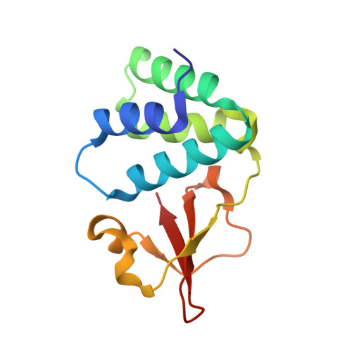 | |
UniProt | |||||
Entity Groups | |||||
| Sequence Clusters | 30% Identity50% Identity70% Identity90% Identity95% Identity100% Identity | ||||
| UniProt Group | Q05127 | ||||
Sequence AnnotationsExpand | |||||
Reference Sequence | |||||
| Length ( Å ) | Angle ( ˚ ) |
|---|---|
| a = 51.75 | α = 90 |
| b = 65.66 | β = 90 |
| c = 72.02 | γ = 90 |
| Software Name | Purpose |
|---|---|
| SCALA | data scaling |
| PHENIX | refinement |
| PDB_EXTRACT | data extraction |
| iMOSFLM | data reduction |
| PHASER | phasing |


