5BPI

Structure of TrmBL2, an archaeal chromatin protein, shows a novel mode of DNA binding.


Experimental Data Snapshot

  • Method: X-RAY DIFFRACTION
  • Resolution: 3.20 Å
  • R-Value Free: 0.299 
  • R-Value Work: 0.255 
  • R-Value Observed: 0.257 

wwPDB Validation   3D Report Full Report


This is version 1.2 of the entry. See complete history


Literature

Structural Insights into Nonspecific Binding of DNA by TrmBL2, an Archaeal Chromatin Protein.

Ahmad, M.U.Waege, I.Hausner, W.Thomm, M.Boos, W.Diederichs, K.Welte, W.

(2015) J Mol Biol 427: 3216-3229

  • DOI: https://doi.org/10.1016/j.jmb.2015.08.012
  • Primary Citation of Related Structures:  
    5BOX, 5BPD, 5BPI, 5BQT

  • PubMed Abstract: 

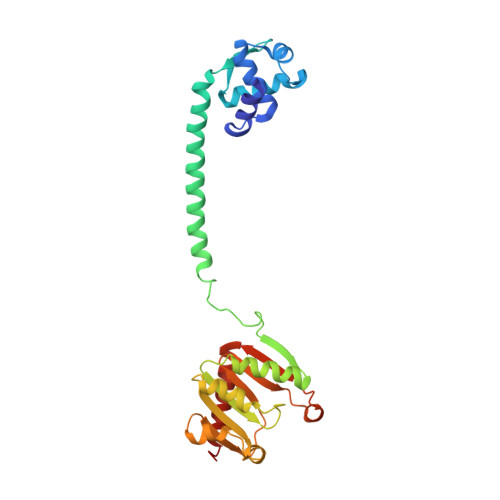
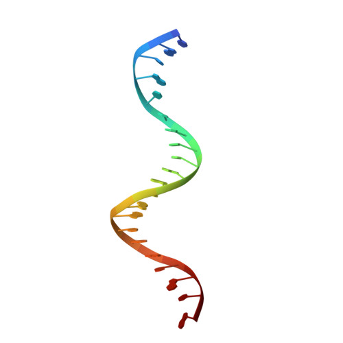
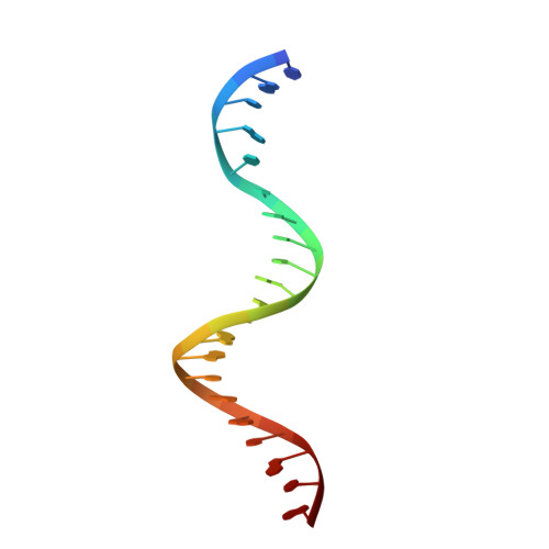
    The crystal structure of TrmBL2 from the archaeon Pyrococcus furiosus shows an association of two pseudosymmetric dimers. The dimers follow the prototypical design of known bacterial repressors with two helix-turn-helix (HTH) domains binding to successive major grooves of the DNA. However, in TrmBL2, the two dimers are arranged at a mutual displacement of approximately 2bp so that they associate with the DNA along the double-helical axis at an angle of approximately 80°. While the deoxyribose phosphate groups of the double-stranded DNA (dsDNA) used for co-crystallization are clearly seen in the electron density map, most of the nucleobases are averaged out. Refinement required to assume a superposition of at least three mutually displaced dsDNAs. The HTH domains interact primarily with the deoxyribose phosphate groups and polar interactions with the nucleobases are almost absent. This hitherto unseen mode of DNA binding by TrmBL2 seems to arise from nonoptimal protein-DNA contacts made by its four HTH domains resulting in a low-affinity, nonspecific binding to DNA.


  • Organizational Affiliation

    University of Konstanz Department of Biology, Universitätsstrasse 10, 78456 Konstanz, Germany.


Macromolecules

Find similar proteins by:  (by identity cutoff)  |  3D Structure
Entity ID: 1
MoleculeChains Sequence LengthOrganismDetailsImage
TrmBL2
A, B, C, D
263Pyrococcus furiosusMutation(s): 0 
Gene Names: trmBL2PF0496
UniProt
Find proteins for Q8U3H1 (Pyrococcus furiosus (strain ATCC 43587 / DSM 3638 / JCM 8422 / Vc1))
Explore Q8U3H1 
Go to UniProtKB:  Q8U3H1
Entity Groups  
Sequence Clusters30% Identity50% Identity70% Identity90% Identity95% Identity100% Identity
UniProt GroupQ8U3H1
Sequence Annotations
Expand
  • Reference Sequence

Find similar nucleic acids by:  Sequence   |   3D Structure  

Entity ID: 2
MoleculeChains LengthOrganismImage
DNA (5'-D(P*TP*AP*TP*AP*TP*CP*AP*TP*CP*GP*AP*TP*AP*GP*TP*GP*AP*TP*AP*TP*A)-3')21Pyrococcus furiosus
Sequence Annotations
Expand
  • Reference Sequence

Find similar nucleic acids by:  Sequence   |   3D Structure  

Entity ID: 3
MoleculeChains LengthOrganismImage
DNA (5'-D(P*TP*AP*TP*AP*TP*CP*AP*CP*TP*AP*TP*CP*GP*AP*TP*GP*AP*TP*AP*TP*A)-3')21Pyrococcus furiosus
Sequence Annotations
Expand
  • Reference Sequence
Small Molecules
Ligands 1 Unique
IDChains Name / Formula / InChI Key2D Diagram3D Interactions
GOL
Query on GOL

Download Ideal Coordinates CCD File 
G [auth A]GLYCEROL
C3 H8 O3
PEDCQBHIVMGVHV-UHFFFAOYSA-N
Experimental Data & Validation

Experimental Data

  • Method: X-RAY DIFFRACTION
  • Resolution: 3.20 Å
  • R-Value Free: 0.299 
  • R-Value Work: 0.255 
  • R-Value Observed: 0.257 
  • Space Group: P 21 21 21
Unit Cell:
Length ( Å )Angle ( ˚ )
a = 58.764α = 90
b = 154.379β = 90
c = 176.123γ = 90
Software Package:
Software NamePurpose
PHENIXrefinement
PDB_EXTRACTdata extraction
XDSdata reduction
XDSdata scaling
Cootmodel building

Structure Validation

View Full Validation Report


Entry History 

Deposition Data

Revision History  (Full details and data files)

  • Version 1.0: 2015-09-02
    Type: Initial release
  • Version 1.1: 2015-10-07
    Changes: Database references
  • Version 1.2: 2024-01-10
    Changes: Data collection, Database references, Refinement description