5BMX | pdb_00005bmx

Crystal structure of T75N mutant of Triosephosphate isomerase from Plasmodium falciparum


Experimental Data Snapshot

  • Method: X-RAY DIFFRACTION
  • Resolution: 1.80 Å
  • R-Value Free: 
    0.216 (Depositor), 0.216 (DCC) 
  • R-Value Work: 
    0.167 (Depositor), 0.167 (DCC) 
  • R-Value Observed: 
    0.169 (Depositor) 

Starting Model: experimental
View more details

wwPDB Validation 3D Report Full Report

Validation slider image for 5BMX

This is version 1.4 of the entry. See complete history

Literature

Probing the role of highly conserved residues in triosephosphate isomerase - analysis of site specific mutants at positions 64 and 75 in the Plasmodial enzyme

Bandyopadhyay, D.Murthy, M.R.Balaram, H.Balaram, P.

(2015) FEBS J 282: 3863-3882

  • DOI: https://doi.org/10.1111/febs.13384
  • Primary Citation Related Structures: 
    4ZZ9, 5BMW, 5BMX, 5BNK, 5BRB

  • PubMed Abstract: 

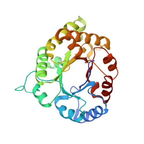
    Highly conserved residues in enzymes are often found to be clustered close to active sites, suggesting that functional constraints dictate the nature of amino acid residues accommodated at these sites. Using the Plasmodium falciparum triosephosphate isomerase (PfTIM) enzyme (EC 5.3.1.1) as a template, we have examined the effects of mutations at positions 64 and 75, which are not directly involved in the proton transfer cycle. Thr (T) occurring at position 75 is completely conserved, whereas only Gln (Q) and Glu (E) are accommodated at position 64. Biophysical and kinetic data are reported for four T75 (T75S/V/C/N) and two Q64 (Q64N/E) mutants. The dimeric structure is weakened in the Q64E and Q64N mutants, whereas dimer integrity is unimpaired in all four T75 mutants. Measurement of the concentration dependence of enzyme activity permits an estimate of Kd values for dimer dissociation (Q64N = 73.7 ± 9.2 nm and Q64E = 44.6 ± 8.4 nm). The T75S/V/C mutants have activities comparable to the wild-type enzyme, whereas a fourfold drop is observed for T75N. All four T75 mutants show a dramatic fall in activity between 35 °C and 45 °C. Crystal structure determination of the T75S/V/N mutants provides insights into the variations in local interactions, with the T75N mutant showing the largest changes. Hydrogen-bond interactions determine dimer stability restricting the choice of residues at position 64 to Gln (Q) and Glu (E). At position 75, the overwhelming preference for Thr (T) may be dictated by the imperative of maintaining temperature stability of enzyme activity. Structural data have been deposited in the Protein Data Bank under accession numbers 4ZZ9, 5BMW, 5BMX, 5BNK and 5BRB.


  • Organizational Affiliation
    • Molecular Biophysics Unit, Indian Institute of Science, Bangalore, India.

Macromolecule Content 

  • Total Structure Weight: 113.02 kDa 
  • Atom Count: 8,651 
  • Modeled Residue Count: 978 
  • Deposited Residue Count: 992 
  • Unique protein chains: 1

Macromolecules

Find similar proteins by:|  3D Structure
Entity ID: 1
MoleculeChains  Sequence LengthOrganismDetailsImage
Triosephosphate isomerase
A, B, C, D
248Plasmodium falciparumMutation(s): 2 
Gene Names: TPI
EC: 5.3.1.1
UniProt
Find proteins for Q07412 (Plasmodium falciparum)
Explore Q07412 
Go to UniProtKB:  Q07412
Entity Groups
Sequence Clusters30% Identity50% Identity70% Identity90% Identity95% Identity100% Identity
UniProt GroupQ07412
Sequence Annotations
Expand
Reference Sequence

Small Molecules

Ligands 3 Unique
IDChains Name / Formula / InChI Key2D Diagram3D Interactions
SO4

Query on SO4


Download:Ideal Coordinates CCD File
H [auth A],
M [auth B],
P [auth C],
T [auth D]
SULFATE ION
O4 S
QAOWNCQODCNURD-UHFFFAOYSA-L
EDO

Query on EDO


Download:Ideal Coordinates CCD File
E [auth A]
F [auth A]
I [auth B]
J [auth B]
K [auth B]
E [auth A],
F [auth A],
I [auth B],
J [auth B],
K [auth B],
N [auth C],
Q [auth D],
R [auth D]
1,2-ETHANEDIOL
C2 H6 O2
LYCAIKOWRPUZTN-UHFFFAOYSA-N
NA

Query on NA


Download:Ideal Coordinates CCD File
G [auth A],
L [auth B],
O [auth C],
S [auth D]
SODIUM ION
Na
FKNQFGJONOIPTF-UHFFFAOYSA-N

Experimental Data & Validation

Experimental Data

  • Method: X-RAY DIFFRACTION
  • Resolution: 1.80 Å
  • R-Value Free:  0.216 (Depositor), 0.216 (DCC) 
  • R-Value Work:  0.167 (Depositor), 0.167 (DCC) 
  • R-Value Observed: 0.169 (Depositor) 
Space Group: P 21 21 21
Unit Cell:
Length ( Å )Angle ( ˚ )
a = 50.64α = 90
b = 106.72β = 90
c = 179.62γ = 90
Software Package:
Software NamePurpose
REFMACrefinement
SCALAdata scaling
PDB_EXTRACTdata extraction
iMOSFLMdata reduction
PHASERphasing

Structure Validation

View Full Validation Report


Entry History 

& Funding Information

Deposition Data


Funding OrganizationLocationGrant Number
Department of BiotechnologyIndia--

Revision History  (Full details and data files)

  • Version 1.0: 2015-07-29
    Type: Initial release
  • Version 1.1: 2015-08-05
    Changes: Database references
  • Version 1.2: 2015-08-12
    Changes: Other
  • Version 1.3: 2015-10-28
    Changes: Database references
  • Version 1.4: 2023-11-08
    Changes: Data collection, Database references, Derived calculations, Refinement description