5B60

Crystal structure of PtLCIB4 S47R mutant, a homolog of the limiting CO2-inducible protein LCIB


Experimental Data Snapshot

  • Method: X-RAY DIFFRACTION
  • Resolution: 2.20 Å
  • R-Value Free: 0.254 
  • R-Value Work: 0.250 
  • R-Value Observed: 0.250 

wwPDB Validation   3D Report Full Report


This is version 1.3 of the entry. See complete history


Literature

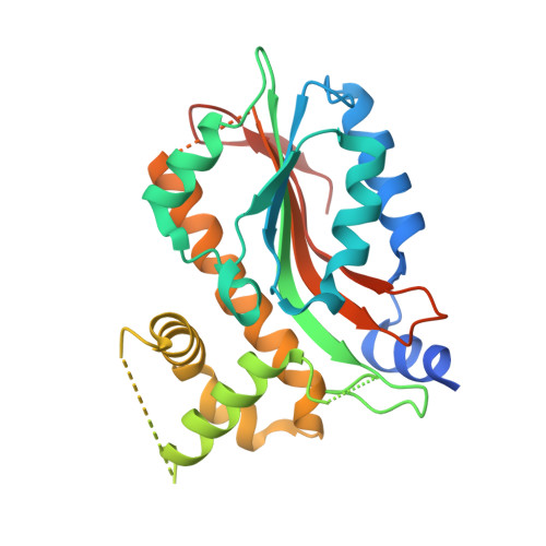
Structural insights into the LCIB protein family reveals a new group of beta-carbonic anhydrases

Jin, S.Sun, J.Wunder, T.Tang, D.Cousins, A.B.Sze, S.K.Mueller-Cajar, O.Gao, Y.G.

(2016) Proc Natl Acad Sci U S A 113: 14716-14721

  • DOI: https://doi.org/10.1073/pnas.1616294113
  • Primary Citation of Related Structures:  
    5B5X, 5B5Y, 5B5Z, 5B60, 5K5W

  • PubMed Abstract: 

    Aquatic microalgae have evolved diverse CO 2 -concentrating mechanisms (CCMs) to saturate the carboxylase with its substrate, to compensate for the slow kinetics and competing oxygenation reaction of the key photosynthetic CO 2 -fixing enzyme rubisco. The limiting CO 2 -inducible B protein (LCIB) is known to be essential for CCM function in Chlamydomonas reinhardtii To assign a function to this previously uncharacterized protein family, we purified and characterized a phylogenetically diverse set of LCIB homologs. Three of the six homologs are functional carbonic anhydrases (CAs). We determined the crystal structures of LCIB and limiting CO 2 -inducible C protein (LCIC) from C. reinhardtii and a CA-functional homolog from Phaeodactylum tricornutum, all of which harbor motifs bearing close resemblance to the active site of canonical β-CAs. Our results identify the LCIB family as a previously unidentified group of β-CAs, and provide a biochemical foundation for their function in the microalgal CCMs.


  • Organizational Affiliation

    School of Biological Sciences, Nanyang Technological University, Singapore 637551.


Macromolecules
Find similar proteins by:  (by identity cutoff)  |  3D Structure
Entity ID: 1
MoleculeChains Sequence LengthOrganismDetailsImage
PtLCIB4 S47R mutant282Phaeodactylum tricornutumMutation(s): 0 
UniProt
Find proteins for B7FR29 (Phaeodactylum tricornutum (strain CCAP 1055/1))
Explore B7FR29 
Go to UniProtKB:  B7FR29
Entity Groups  
Sequence Clusters30% Identity50% Identity70% Identity90% Identity95% Identity100% Identity
UniProt GroupB7FR29
Sequence Annotations
Expand
  • Reference Sequence
Experimental Data & Validation

Experimental Data

  • Method: X-RAY DIFFRACTION
  • Resolution: 2.20 Å
  • R-Value Free: 0.254 
  • R-Value Work: 0.250 
  • R-Value Observed: 0.250 
  • Space Group: C 1 2 1
Unit Cell:
Length ( Å )Angle ( ˚ )
a = 68.55α = 90
b = 66.11β = 101
c = 69.06γ = 90
Software Package:
Software NamePurpose
PHENIXrefinement
SCALAdata reduction
SCALAdata scaling
PHENIXphasing

Structure Validation

View Full Validation Report


Entry History & Funding Information

Deposition Data


Funding OrganizationLocationGrant Number
Singapore National Research FoundationSingaporeNRF-RF2009-RF001-267

Revision History  (Full details and data files)

  • Version 1.0: 2016-12-07
    Type: Initial release
  • Version 1.1: 2017-01-18
    Changes: Database references
  • Version 1.2: 2020-02-26
    Changes: Data collection
  • Version 1.3: 2023-11-08
    Changes: Data collection, Database references, Refinement description