5B4I

Crystal structure of I86D mutant of phycocyanobilin:ferredoxin oxidoreductase in complex with biliverdin (data 2)


Experimental Data Snapshot

  • Method: X-RAY DIFFRACTION
  • Resolution: 1.11 Å
  • R-Value Free: 0.152 
  • R-Value Work: 0.127 

wwPDB Validation   3D Report Full Report


Ligand Structure Quality Assessment 


This is version 1.2 of the entry. See complete history


Literature

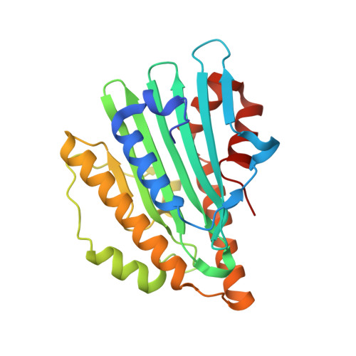
Atomic-resolution structure of the phycocyanobilin:ferredoxin oxidoreductase I86D mutant in complex with fully protonated biliverdin

Hagiwara, Y.Wada, K.Irikawa, T.Sato, H.Unno, M.Yamamoto, K.Fukuyama, K.Sugishima, M.

(2016) FEBS Lett 590: 3425-3434

  • DOI: https://doi.org/10.1002/1873-3468.12387
  • Primary Citation of Related Structures:  
    5B4H, 5B4I, 5B4J

  • PubMed Abstract: 

    Phycocyanobilin:ferredoxin oxidoreductase (PcyA) catalyzes the reduction of biliverdin (BV) to produce phycocyanobilin, a linear tetrapyrrole pigment used for light harvesting and light sensing. Spectroscopic and HPLC analyses inidicate that BV bound to the I86D mutant of PcyA is fully protonated (BVH + ) and can accept an electron, but I86D is unable to donate protons for the reduction; therefore, compared to the wild-type PcyA, the I86D mutant stabilizes BVH + . To elucidate the structural basis of the I86D mutation, we determined the atomic-resolution structure of the I86D-BVH + complex and the protonation states of the essential residues Asp105 and Glu76 in PcyA. Our study revealed that Asp105 adopted a fixed conformation in the I86D mutant, although it had dual conformations in wild-type PcyA which reflected the protonation states of BV. Taken together with biochemical/spectroscopic results, our analysis of the I86D-BVH + structure supports the hypothesis that flexibility of Asp105 is essential for the catalytic activity of PcyA.


  • Organizational Affiliation

    Department of Biochemistry and Applied Chemistry, National Institute of Technology, Kurume College, Kurume, Japan.


Macromolecules
Find similar proteins by:  (by identity cutoff)  |  3D Structure
Entity ID: 1
MoleculeChains Sequence LengthOrganismDetailsImage
Phycocyanobilin:ferredoxin oxidoreductase248Synechocystis sp. PCC 6803Mutation(s): 1 
Gene Names: pcyA
EC: 1.3.7.5
UniProt
Find proteins for Q55891 (Synechocystis sp. (strain PCC 6803 / Kazusa))
Explore Q55891 
Go to UniProtKB:  Q55891
Entity Groups  
Sequence Clusters30% Identity50% Identity70% Identity90% Identity95% Identity100% Identity
UniProt GroupQ55891
Sequence Annotations
Expand
  • Reference Sequence
Small Molecules
Ligands 1 Unique
IDChains Name / Formula / InChI Key2D Diagram3D Interactions
BLA
Query on BLA

Download Ideal Coordinates CCD File 
B [auth A]BILIVERDINE IX ALPHA
C33 H34 N4 O6
GWZYPXHJIZCRAJ-SRVCBVSDSA-N
Experimental Data & Validation

Experimental Data

  • Method: X-RAY DIFFRACTION
  • Resolution: 1.11 Å
  • R-Value Free: 0.152 
  • R-Value Work: 0.127 
  • Space Group: P 21 21 2
Unit Cell:
Length ( Å )Angle ( ˚ )
a = 70.833α = 90
b = 95.691β = 90
c = 42.398γ = 90
Software Package:
Software NamePurpose
SHELXL-97refinement
PHENIXphasing
HKL-2000data reduction
HKL-2000data scaling
SHELXphasing

Structure Validation

View Full Validation Report


Ligand Structure Quality Assessment 


Entry History & Funding Information

Deposition Data


Funding OrganizationLocationGrant Number
JSPSJapan20370037
JSPSJapan22770096
JSPSJapan23370052
JSPSJapan24570122
JSPSJapan24870030
JSPSJapan25840026
MEXT, Program to Disseminate Tenura TrackingJapan--

Revision History  (Full details and data files)

  • Version 1.0: 2017-03-15
    Type: Initial release
  • Version 1.1: 2020-02-26
    Changes: Data collection
  • Version 1.2: 2023-11-08
    Changes: Data collection, Database references, Refinement description