Angucycline antibiotic waldiomycin recognizes common structural motif conserved in bacterial histidine kinases
Eguchi, Y., Okajima, T., Tochio, N., Inukai, Y., Shimizu, R., Ueda, S., Shinya, S., Kigawa, T., Fukamizo, T., Igarashi, M., Utsumi, R.(2017) J Antibiot (Tokyo) 70: 251-258
- PubMed: 27999439 Search on PubMed
- DOI: https://doi.org/10.1038/ja.2016.151
- Primary Citation Related Structures:
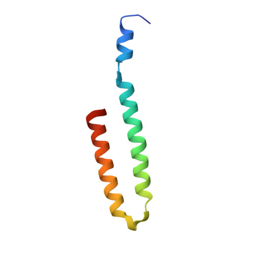
5B1N, 5B1O - PubMed Abstract:
Two-component signal transduction systems (TCSs), composed of a histidine kinase sensor (HK) and its cognate response regulator, sense and respond to environmental changes and are related to the virulence of pathogens. TCSs are potential targets for alternative antibiotics and anti-virulence agents. Here we found that waldiomycin, an angucycline antibiotic that inhibits a growth essential HK, WalK, in Gram-positive bacteria, also inhibits several class I HKs from the Gram-negative Escherichia coli. NMR analyses and site-directed mutagenesis studies using the osmo-sensing EnvZ, a prototypical HK of E. coli, showed that waldiomycin directly binds to both H-box and X-region, which are the two conserved regions in the dimerization-inducing and histidine-containing phosphotransfer (DHp) domain of HKs. Waldiomycin inhibits phosphorylation of the conserved histidine in the H-box. Analysis of waldiomycin derivatives suggests that the angucyclic ring, situated near the H-box in the waldiomycin-EnvZ DHp domain complex model, is responsible for the inhibitory activity. We demonstrate that waldiomycin is an HK inhibitor binding to the H-box region and has the potential of inhibiting a broad spectrum of HKs.
- Department of Bioscience, Graduate School of Agriculture, Kindai University, Nara, Japan.
Organizational Affiliation: