5AWX | pdb_00005awx

Crystal structure of Human PTPRZ D1 domain


Experimental Data Snapshot

  • Method: X-RAY DIFFRACTION
  • Resolution: 1.86 Å
  • R-Value Free: 
    0.221 (Depositor), 0.223 (DCC) 
  • R-Value Work: 
    0.186 (Depositor), 0.188 (DCC) 
  • R-Value Observed: 
    0.187 (Depositor) 

Starting Model: experimental
View more details

wwPDB Validation 3D Report Full Report

Validation slider image for 5AWX

This is version 1.2 of the entry. See complete history

Literature

Small-molecule inhibition of PTPRZ reduces tumor growth in a rat model of glioblastoma

Fujikawa, A.Nagahira, A.Sugawara, H.Ishii, K.Imajo, S.Matsumoto, M.Kuboyama, K.Suzuki, R.Tanga, N.Noda, M.Uchiyama, S.Tomoo, T.Ogata, A.Masumura, M.Noda, M.

(2016) Sci Rep 6: 20473-20473

  • DOI: https://doi.org/10.1038/srep20473
  • Primary Citation Related Structures: 
    5AWX

  • PubMed Abstract: 

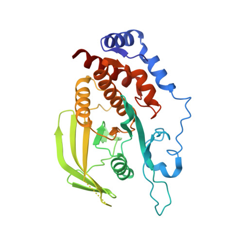
    Protein tyrosine phosphatase receptor-type Z (PTPRZ) is aberrantly over-expressed in glioblastoma and a causative factor for its malignancy. However, small molecules that selectively inhibit the catalytic activity of PTPRZ have not been discovered. We herein performed an in vitro screening of a chemical library, and identified SCB4380 as the first potent inhibitor for PTPRZ. The stoichiometric binding of SCB4380 to the catalytic pocket was demonstrated by biochemical and mass spectrometric analyses. We determined the crystal structure of the catalytic domain of PTPRZ, and the structural basis of the binding of SCB4380 elucidated by a molecular docking method was validated by site-directed mutagenesis studies. The intracellular delivery of SCB4380 by liposome carriers inhibited PTPRZ activity in C6 glioblastoma cells, and thereby suppressed their migration and proliferation in vitro and tumor growth in a rat allograft model. Therefore, selective inhibition of PTPRZ represents a promising approach for glioma therapy.


  • Organizational Affiliation
    • Division of Molecular Neurobiology, National Institute for Basic Biology (NIBB), 5-1 Higashiyama, Myodaiji-cho, Okazaki, Aichi, 444-8787, Japan.

Macromolecule Content 

  • Total Structure Weight: 34.27 kDa 
  • Atom Count: 2,540 
  • Modeled Residue Count: 283 
  • Deposited Residue Count: 298 
  • Unique protein chains: 1

Macromolecules

Find similar proteins by:|  3D Structure
Entity ID: 1
MoleculeChains  Sequence LengthOrganismDetailsImage
Receptor-type tyrosine-protein phosphatase zeta298Homo sapiensMutation(s): 0 
Gene Names: PTPRZ1HTPZP2PTPRZPTPRZ2PTPZ
EC: 3.1.3.48
UniProt & NIH Common Fund Data Resources
Find proteins for P23471 (Homo sapiens)
Explore P23471 
Go to UniProtKB:  P23471
PHAROS:  P23471
GTEx:  ENSG00000106278 
Entity Groups
Sequence Clusters30% Identity50% Identity70% Identity90% Identity95% Identity100% Identity
UniProt GroupP23471
Sequence Annotations
Expand
Reference Sequence

Experimental Data & Validation

Experimental Data

  • Method: X-RAY DIFFRACTION
  • Resolution: 1.86 Å
  • R-Value Free:  0.221 (Depositor), 0.223 (DCC) 
  • R-Value Work:  0.186 (Depositor), 0.188 (DCC) 
  • R-Value Observed: 0.187 (Depositor) 
Space Group: P 21 21 21
Unit Cell:
Length ( Å )Angle ( ˚ )
a = 54.641α = 90
b = 72.245β = 90
c = 90.557γ = 90
Software Package:
Software NamePurpose
REFMACrefinement
HKL-2000data reduction
HKL-2000data scaling
AMoREphasing

Structure Validation

View Full Validation Report


Entry History 

Deposition Data

  • Released Date: 2016-02-17 
  • Deposition Author(s): Sugawara, H.

Revision History  (Full details and data files)

  • Version 1.0: 2016-02-17
    Type: Initial release
  • Version 1.1: 2016-03-09
    Changes: Database references
  • Version 1.2: 2023-11-08
    Changes: Data collection, Database references, Derived calculations, Refinement description