


Crystal Structure and DNA Binding Ability of Myco Tuberculosis Vapbc49 Anti-Toxin Protein Bacterium
Holton, S.J., Pogenberg, V., Iborra, V., Wilmanns, M.To be published.
Experimental Data Snapshot
wwPDB Validation 3D Report Full Report

Entity ID: 1 | |||||
|---|---|---|---|---|---|
| Molecule | Chains | Sequence Length | Organism | Details | Image |
| VAPBC49 | 241 | Mycobacterium tuberculosis H37Rv | Mutation(s): 0 | 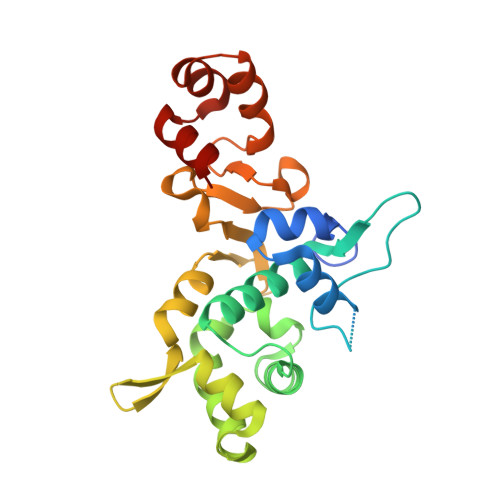 | |
UniProt | |||||
Entity Groups | |||||
| Sequence Clusters | 30% Identity50% Identity70% Identity90% Identity95% Identity100% Identity | ||||
| UniProt Group | O53464 | ||||
Sequence AnnotationsExpand | |||||
Reference Sequence | |||||
| Ligands 1 Unique | |||||
|---|---|---|---|---|---|
| ID | Chains | Name / Formula / InChI Key | 2D Diagram | 3D Interactions | |
| GOL Download:Ideal Coordinates CCD File | C [auth A] | GLYCEROL C3 H8 O3 PEDCQBHIVMGVHV-UHFFFAOYSA-N |  | ||
| Length ( Å ) | Angle ( ˚ ) |
|---|---|
| a = 81.935 | α = 90 |
| b = 81.935 | β = 90 |
| c = 75.361 | γ = 120 |
| Software Name | Purpose |
|---|---|
| REFMAC | refinement |


