5A81 | pdb_00005a81

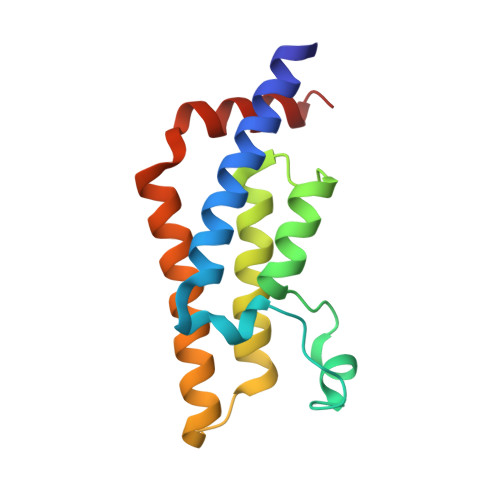
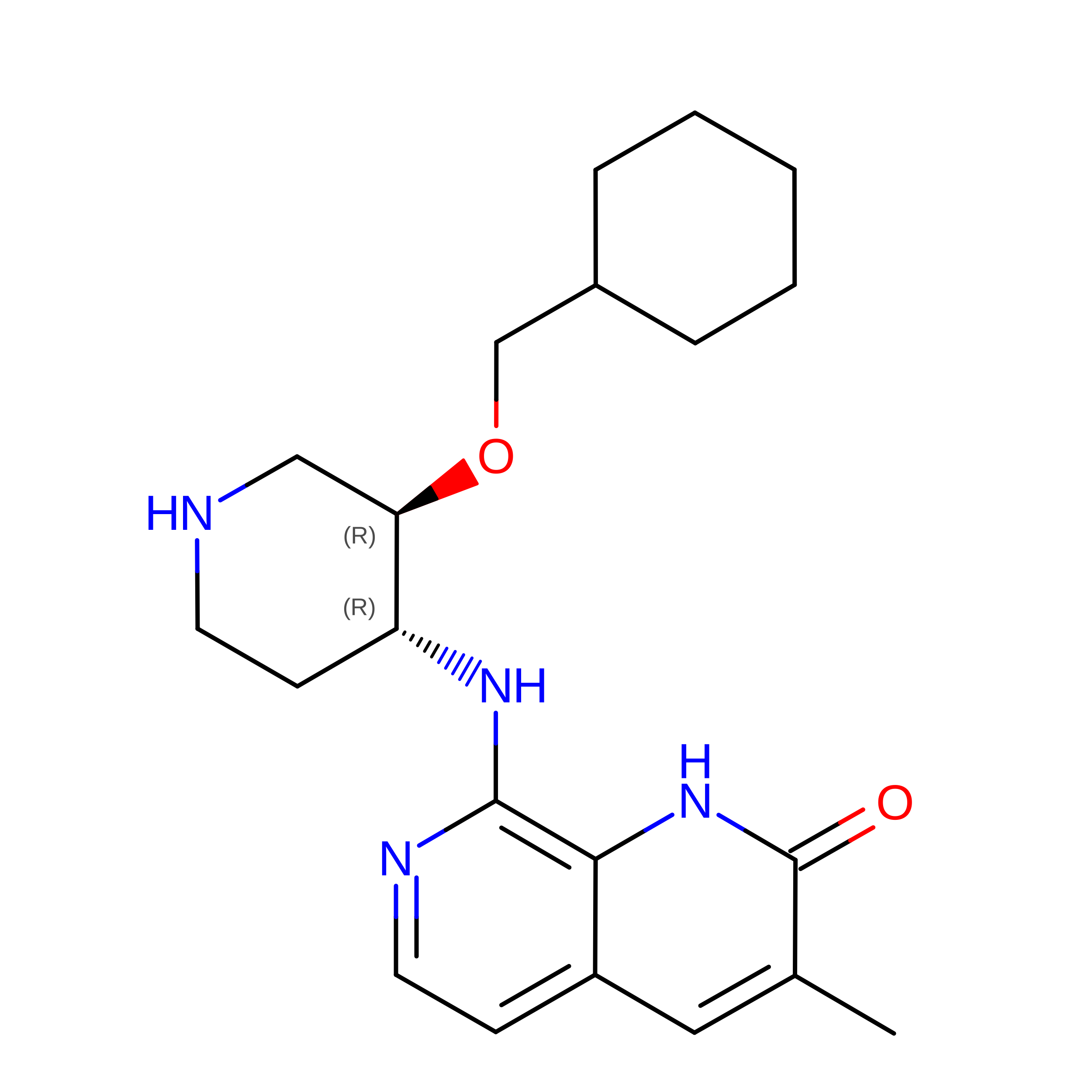
Crystal structure of human ATAD2 bromodomain in complex with 8-(3R,4R) -3-(cyclohexylmethoxy)piperidin-4-yl-amino-3-methyl-1,2-dihydro-1,7- naphthyridin-2-one


Experimental Data Snapshot

  • Method: X-RAY DIFFRACTION
  • Resolution: 2.03 Å
  • R-Value Free: 
    0.219 (Depositor), 0.209 (DCC) 
  • R-Value Work: 
    0.197 (Depositor) 
  • R-Value Observed: 
    0.198 (Depositor) 

wwPDB Validation 3D Report Full Report

Validation slider image for 5A81

Ligand Structure Quality Assessment 


This is version 1.2 of the entry. See complete history. 

Literature

Structure-Based Optimization of Naphthyridones Into Potent Atad2 Bromodomain Inhibitors.

Bamborough, P., Chung, C., Furze, R.C., Grandi, P., Michon, A., Sheppard, R.J., Barnett, H., Diallo, H., Dixon, D.P., Douault, C., Jones, E.J., Karamshi, B., Mitchell, D.J., Prinjha, R.K., Rau, C., Watson, R.J., Werner, T., Demont, E.H.

(2015) J Med Chem 58: 6151

  • DOI: https://doi.org/10.1021/acs.jmedchem.5b00773
  • Primary Citation Related Structures: 
    5A81, 5A82, 5A83, 5A85

  • PubMed Abstract: 

    ATAD2 is a bromodomain-containing protein whose overexpression is linked to poor outcomes in a number of different cancer types. To date, no potent and selective inhibitors of the bromodomain have been reported. This article describes the structure-based optimization of a series of naphthyridones from micromolar leads with no selectivity over the BET bromodomains to inhibitors with sub-100 nM ATAD2 potency and 100-fold BET selectivity.


  • Organizational Affiliation: 
    • ∥Cellzome GmbH, Molecular Discovery Research, GlaxoSmithKline, Meyerhofstrasse 1, 69117 Heidelberg, Germany.

Macromolecule Content 

  • Total Structure Weight: 16.14 kDa 
  • Atom Count: 1,349 
  • Modeled Residue Count: 130 
  • Deposited Residue Count: 130 
  • Unique protein chains: 1

Macromolecules

Find similar proteins by:|  3D Structure
Entity ID: 1
MoleculeChains  Sequence LengthOrganismDetailsImage
ATPASE FAMILY AAA DOMAIN-CONTAINING PROTEIN 2130Homo sapiensMutation(s): 0 
EC: 3.6.1.3 (PDB Primary Data), 3.6.1 (UniProt)
UniProt & NIH Common Fund Data Resources
Find proteins for Q6PL18 (Homo sapiens)
Explore Q6PL18 
Go to UniProtKB:  Q6PL18
PHAROS:  Q6PL18
GTEx:  ENSG00000156802 
Entity Groups
Sequence Clusters30% Identity50% Identity70% Identity90% Identity95% Identity100% Identity
UniProt GroupQ6PL18
Sequence Annotations
Expand
Reference Sequence

Small Molecules

Binding Affinity Annotations 
IDSourceBinding Affinity
78J BindingDB:  5A81 Kd: min: 2900, max: 3162 (nM) from 3 assay(s)
IC50: min: 2512, max: 3981 (nM) from 4 assay(s)

Experimental Data & Validation

Experimental Data

  • Method: X-RAY DIFFRACTION
  • Resolution: 2.03 Å
  • R-Value Free:  0.219 (Depositor), 0.209 (DCC) 
  • R-Value Work:  0.197 (Depositor) 
  • R-Value Observed: 0.198 (Depositor) 
Space Group: P 65 2 2
Unit Cell:
Length ( Å )Angle ( ˚ )
a = 79.54α = 90
b = 79.54β = 90
c = 138.22γ = 120
Software Package:
Software NamePurpose
REFMACrefinement
XDSdata reduction
Aimlessdata scaling

Structure Validation

View Full Validation Report


Ligand Structure Quality Assessment 


Entry History 

Deposition Data

Revision History  (Full details and data files)

  • Version 1.0: 2015-08-12
    Type: Initial release
  • Version 1.1: 2015-08-26
    Changes: Database references
  • Version 1.2: 2024-05-08
    Changes: Data collection, Database references, Derived calculations, Other