Crystal Structure of Arabidopsis Thaliana Calmodulin7 and Insight Into its Mode of DNA Binding.
Kumar, S., Mazumder, M., Gupta, N., Chattopadhyay, S., Gourinath, S.(2016) FEBS Lett 590: 3029
- PubMed: 27500568 Search on PubMed
- DOI: https://doi.org/10.1002/1873-3468.12349
- Primary Citation Related Structures:
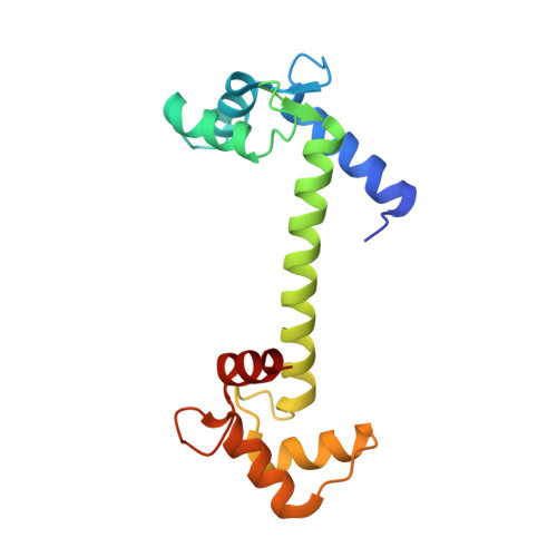
5A2H - PubMed Abstract:
Calmodulin (CaM) is a Ca(2+) sensor that participates in several cellular signaling cascades by interacting with various targets, including DNA. It has been shown that Arabidopsis thaliana CaM7 (AtCaM7) interacts with Z-box DNA and functions as a transcription factor [Kushwaha R et al. (2008) Plant Cell 20, 1747-1759; Abbas N et al. (2014) Plant Cell 26, 1036-1052]. The crystal structure of AtCaM7, and a model of the AtCAM7-Z-box complex suggest that Arg-127 determines the DNA-binding ability by forming crucial interactions with the guanine base. We validated the model using biolayer interferometry, which confirmed that AtCaM7 interacts with Z-box DNA with high affinity. In contrast, the AtCaM2/3/5 isoform does not show any binding, although it differs from AtCaM7 by only a single residue.
- School of Life Sciences, Jawaharlal Nehru University, New Delhi, India.
Organizational Affiliation: