5A1S | pdb_00005a1s

Crystal structure of the sodium-dependent citrate symporter SeCitS form Salmonella enterica.


Experimental Data Snapshot

  • Method: X-RAY DIFFRACTION
  • Resolution: 2.50 Å
  • R-Value Free: 
    0.246 (Depositor), 0.247 (DCC) 
  • R-Value Work: 
    0.208 (Depositor), 0.212 (DCC) 
  • R-Value Observed: 
    0.210 (Depositor) 

wwPDB Validation 3D Report Full Report

Validation slider image for 5A1S

Ligand Structure Quality Assessment 


This is version 1.3 of the entry. See complete history

Literature

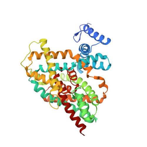
Mechanism of Na(+)-dependent citrate transport from the structure of an asymmetrical CitS dimer.

Wohlert, D.Grotzinger, M.J.Kuhlbrandt, W.Yildiz, O.

(2015) Elife 4: e09375-e09375

  • DOI: https://doi.org/10.7554/eLife.09375
  • Primary Citation Related Structures: 
    5A1S

  • PubMed Abstract: 

    The common human pathogen Salmonella enterica takes up citrate as a nutrient via the sodium symporter SeCitS. Uniquely, our 2.5 Å x-ray structure of the SeCitS dimer shows three different conformations of the active protomer. One protomer is in the outside-facing state. Two are in different inside-facing states. All three states resolve the substrates in their respective binding environments. Together with comprehensive functional studies on reconstituted proteoliposomes, the structures explain the transport mechanism in detail. Our results indicate a six-step process, with a rigid-body 31° rotation of a helix bundle that translocates the bound substrates by 16 Å across the membrane. Similar transport mechanisms may apply to a wide variety of related and unrelated secondary transporters, including important drug targets.


  • Organizational Affiliation
    • Department of Structural Biology, Max Planck Institute of Biophysics, Frankfurt am Main, Germany.

Macromolecule Content 

  • Total Structure Weight: 195.95 kDa 
  • Atom Count: 13,270 
  • Modeled Residue Count: 1,721 
  • Deposited Residue Count: 1,792 
  • Unique protein chains: 1

Macromolecules

Find similar proteins by:|  3D Structure
Entity ID: 1
MoleculeChains  Sequence LengthOrganismDetailsImage
CITRATE-SODIUM SYMPORTER
A, B, C, D
448Salmonella entericaMutation(s): 0 

Small Molecules

Ligands 7 Unique
IDChains Name / Formula / InChI Key2D Diagram3D Interactions
PTY

Query on PTY


Download:Ideal Coordinates CCD File
M [auth A]PHOSPHATIDYLETHANOLAMINE
C40 H80 N O8 P
NJGIRBISCGPRPF-KXQOOQHDSA-N
BOG

Query on BOG


Download:Ideal Coordinates CCD File
DA [auth D],
H [auth A],
N [auth A],
U [auth C],
V [auth C]
octyl beta-D-glucopyranoside
C14 H28 O6
HEGSGKPQLMEBJL-RKQHYHRCSA-N
MES

Query on MES


Download:Ideal Coordinates CCD File
CA [auth D]2-(N-MORPHOLINO)-ETHANESULFONIC ACID
C6 H13 N O4 S
SXGZJKUKBWWHRA-UHFFFAOYSA-N
FLC

Query on FLC


Download:Ideal Coordinates CCD File
G [auth A]
K [auth A]
L [auth A]
P [auth B]
Q [auth B]
G [auth A],
K [auth A],
L [auth A],
P [auth B],
Q [auth B],
T [auth C],
Y [auth D]
CITRATE ANION
C6 H5 O7
KRKNYBCHXYNGOX-UHFFFAOYSA-K
UND

Query on UND


Download:Ideal Coordinates CCD File
I [auth A],
Z [auth D]
UNDECANE
C11 H24
RSJKGSCJYJTIGS-UHFFFAOYSA-N
CL

Query on CL


Download:Ideal Coordinates CCD File
AA [auth D],
BA [auth D],
J [auth A]
CHLORIDE ION
Cl
VEXZGXHMUGYJMC-UHFFFAOYSA-M
NA

Query on NA


Download:Ideal Coordinates CCD File
E [auth A]
F [auth A]
O [auth B]
R [auth C]
S [auth C]
E [auth A],
F [auth A],
O [auth B],
R [auth C],
S [auth C],
W [auth D],
X [auth D]
SODIUM ION
Na
FKNQFGJONOIPTF-UHFFFAOYSA-N

Experimental Data & Validation

Experimental Data

  • Method: X-RAY DIFFRACTION
  • Resolution: 2.50 Å
  • R-Value Free:  0.246 (Depositor), 0.247 (DCC) 
  • R-Value Work:  0.208 (Depositor), 0.212 (DCC) 
  • R-Value Observed: 0.210 (Depositor) 
Space Group: P 1
Unit Cell:
Length ( Å )Angle ( ˚ )
a = 86.382α = 90.44
b = 89.935β = 113.79
c = 91.843γ = 99.55
Software Package:
Software NamePurpose
PARROTmodel building
Aimlessdata scaling
SHELXDphasing
CRANK2phasing
PARROTphasing
PHENIXrefinement

Structure Validation

View Full Validation Report


Ligand Structure Quality Assessment 


Entry History 

Deposition Data

Revision History  (Full details and data files)

  • Version 1.0: 2016-02-17
    Type: Initial release
  • Version 1.1: 2017-03-22
    Changes: Database references
  • Version 1.2: 2020-07-29
    Type: Remediation
    Reason: Carbohydrate remediation
    Changes: Data collection, Derived calculations, Other, Structure summary
  • Version 1.3: 2024-05-08
    Changes: Data collection, Database references, Structure summary